What is Dual Source Dual Energy?


Dual Source Dual Energy

While the concept of Dual Energy CT was introduced by Siemens more than 30 years ago with fast kV switching on the SOMATOM DRH1, it wasn’t until about 10 years ago that Siemens Healthineers was able to bring Dual Source Technology to the clinical market and with it a more advanced way of acquiring spectral data - Dual Source Dual Energy. This advance made it realistic to utilize Dual Energy in the clinical routine by enabling a method that is able to use modern dose reduction technologies such iterative reconstruction and automated exposure control (dose modulation) while also bringing new ideas such as spectral shaping with tin filtration for acquiring spectral data in a dose neutral manner2 without a loss of image quality that occurs with fast kV switching.

Dual Source Dual Energy

“… virtual monoenergetic reconstruction at 55 keV in pancreatic DECT showed improved objective image quality and reader preference compared to routine images. As this image reconstruction can be incorporated into the scan protocol, this technique should be considered for routine clinical use.”3
 

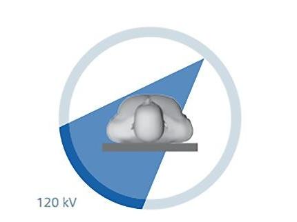
The launch of the SOMATOM Force with its Selective Photon Shield further established Dual Energy as a dose-neutral technology. The Selective Photon Shield filters out unnecessary photons from the high-energy X-ray tube. It permits the use of Dual Energy in daily practice at a dose comparable to a conventional 120-kV scan, with optimal spectra separation for excellent Dual Energy data quality.


Siemens Healthineers Dual Source Dual Energy offers three main advantages: High power, High speed and personalization. These three attributes translate into better image quality for nearly every possible type of patient, images offering you the potential for greater clinical accuracy and diagnostic confidence, and the ability to tailor your dose and scan protocols for even the most challenging scans. By facilitating optimum image quality even in most challenging cases across all medical fields, Dual Source CT has multiplied the potential of Computed Tomography – regarding both application range and information quality.


In 10 years, in over 1,500 installations around the world, and in thousands of clinical cases, Dual Source scanning has proven a great advantage for clinical routines across all fields of medical imaging.
 

In contrast to traditional single source multi-slice CT (Fig 1), a Dual Source CT (DSCT) is equipped with two data measurement systems, each consisting of one X-ray tube and one corresponding detector array oriented in the gantry with an angular offset of 90 degrees (Fig. 2).
 

Dual Source Dual Energy
Fig. 1: Conventional multislice CT equipped with one X-ray generator

The two X-ray source/detector systems rotate simultaneously capturing image data in half the time required with conventional technology. With Dual Source CT it is possible to double the temporal resolution to 66 ms compared with that of a single source CT, and increase the speed of acquisition while reducing the radiation dose significantly.

With the advantage of two measurement systems, DSCT offers a subset of unique advantages including two unique operating modes: Dual Source Single Energy (DSSE) and Dual Source Dual Energy (DSDE) modes.

In DSSE mode, both X-ray tubes work at the same kVp setting and provide extremely fast volumetric coverage (e.g. Flash/Turbo Flash modes) providing both the power and speed for imaging very obese patients (combining the power of two tubes), whole body trauma and cardiac imaging. Learn more about DSCT click here.

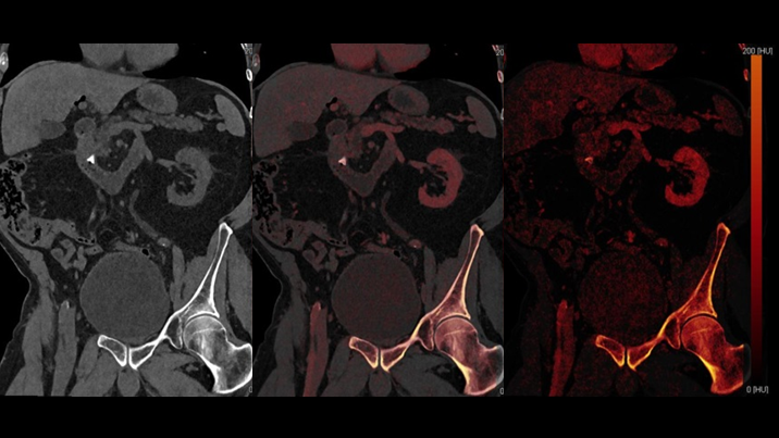
In DSDE mode, X-ray tubes are set at different energies (see Fig. 3) to provide the maximum spectral separation, which is the key to high sensitivity and specificity in spectral imaging, customized for each patient and each acquisition. Dual energy spectral data provides added insight over traditional structural only images by making it possible to differentiate not only between fat, soft tissue, and bone, but also between calcifications and contrast material (iodine) on the basis of their unique energy-dependent attenuation profiles. Furthermore, functional parameters such as iodine concentration in the liver, lung, myocardium or tumors etc. can be acquired. DSDE also enables automatic zero-click bone removal in angiography in both the head and body.

Dual Source Dual Energy
Fig. 3: Dual Source Dual Energy imaging optimizes dose performance and spectral separation by simultaneously with two X-ray sources at different energy levels.

Power


Dual Source Dual Energy

Approximately 65% of US adults are either overweight or obese[1]. This presents a challenge to acquire suitable clinical images, even with the most advanced single source scanners. Dual source scanners offer up to 2600mA to handle your biggest patients. Diagnostic results regardless of a patient’s age, size, weight, physical condition, and even the surrounding circumstances directly translates into more informed decisions, and thus, into more promising patient outcomes.


Not all CT scanners are built to the same standard. Our Dual Source scanners are industry leaders with two powerful generators (up to 120kW) tuned to provide x-ray output in 10 keV steps. This enables you to offer the optimal kVp pair combination with the greatest spectral separation for each patient and scan acquisition. This is all done while offering you up to 1300 mA on each tube, an unheard of level in the industry, providing the power you need to scan patients of any size or makeup. When using other DE approaches to scan bariatric patients, image quality and spectral precision is sacrificed due to system limitations, with Dual Source Dual Energy (DSDE) you have the right balance to ensure high quality images. More importantly without providing a high dose in return 4,5,6

Dual Source Dual Energy
*Bariatric Image courtesy of Northwestern University, Illinois

425 lb patient with a Pancreatic mass that is difficult to identify on a mixed 120 kV image, but using the Monoenergetic Plus application, the mass becomes clearly visualized. Why limit your diagnosis to one way of reading the images?

The launch of the SOMATOM ® Force with its Selective Photon Shield II further established Dual Energy as a dose-neutral technology. The Selective Photon Shield filters out unnecessary photons from the high-energy X-ray tube. It permits the use of Dual Energy in daily practice at a dose comparable to a conventional 120-kV scan, with optimal spectra separation for excellent Dual Energy data quality. Up to 30% increased energy separation for better DE imaging outcomes.

Dual Source Dual Energy

“… virtual monoenergetic reconstruction at 55 keV in pancreatic DECT showed improved objective image quality and reader preference compared to routine images. As this image reconstruction can be incorporated into the scan protocol, this technique should be considered for routine clinical use.” 3

Dual Source Dual Energy

Precise DE quantification – tissue differentiation and therapy response assessment of liver cancer

Dose neutral Dual Energy, through improved iodine visualization and quantification aids in proper physician evaluation of liver lesions.

*Courtesy of University Hospital Zurich, Switzerland
 

Speed


Fast scan speeds supporting your entire patient load

Uncompromised speed to handle the emergency patients

Reliable, CT-based diagnosis in acute care? Within seconds? Maximum possible confidence when time is tight? Dual Source Dual Energy CT imaging without any compromises.

Data7 shows that ~74% of all acquisitions are greater than 16cm in length. Particularly in trauma cases, you may not know the full extent of injuries before CT acquisition thus making it necessary to examine larger extents of the body. Multi-spectral CT data can provide additional information to aid in critical diagnosis and you wouldn’t want to sacrifice this benefit for patients who are compromised and cannot stay still, speed is a required during multi-spectral CT acquisition. With industry leading acquisition speed, DSDE scanners are able to handle all your patients, from “typical” to “critical”. In cases of severe trauma, especially when internal bleeding is involved, complications such as shock may occur if the patient is not managed appropriately and efficiently. Speed is THE priority. Dual Source Dual Energy can help you find the right answers in less time realizing the full benefits of multi-spectral CT.
DSDE can provide: efficiency improvements with full body bone removal helping you to identify abnormalities in vascular exams; identification of bone marrow edema in acute fracture, fat emboli attributable to fracture and obscure vascular obstructions like bowel ischemia; improved conspicuity of active/acute hemorrhage while making it easier to differentiate it from calcium ; aid the diagnosis of brain hemorrhage through accurate quantification and visualization of iodine.


Speed
*Image Courtesy of Brigham and Women's Hospital

DSDE has the speed for ED and trauma patients. When you scan patients presenting with abdominal pain, with DSDE acquiring multi-spectral CT can be your normal workflow, helping you to identify obscure abnormalities such as the bowel ischemia.

Calcium Suppression for the Visualization of Bone Marrow Edema in Acute Fracture
*Image Courtesy of the Massachusetts General Hospital

One click color-coding of bone marrow to evaluate bone bruises and diffuse tumor infiltrations.
 

Precise Dual Energy tissue differentiation – cardiac PBV stenosis
* Image Courtesy of MUSC Medical Center/Charleston, USA

Dual Source delivers detailed, diagnostic cardiac images for all heart rates. When combined with dual energy, DSDE maintains the ultra-fast 66ms temporal resolution for structural imaging and allow for precise iodine quantification in the heart parenchyma.
Advanced diagnostic information due to Dual Energy. With one scan, both CTA and myocardium PBV (Perfusion Blood Volume) information are acquired without any dose penalty for dual energy acquisition.
 

SN

[The most consequential benefit of Dual Source is] Improving patient outcomes and helping us make life saving decisions. The machine delivers diagnostic imaging needed to help us save lives. I truly believe that in the acute setting. We see it day in and day out. To be able to make critical decisions in the matter of seconds, where it matters most.

Savvas Nicolaou, MD,
Associate Professor of Diagnostic Radiology,
Vice-Chair of the Undergraduate Education and Continuing Professional Development for the UBC Department of Radiology,
Head of Emergency and Trauma Radiology at Vancouver General Hospital, Vancouver, Canada

Personalization


Uncompromised personalization to offer the best image quality to every patient

Uncompromised personalization to offer the best image quality to every patient

Efficiency is critical. Determining whether or not you can utilize dual energy for every patient is cumbersome and time consuming - adding complexity to your workflow. By retaining dose modulation on Dual Source Dual Energy, the greater insight from multi-spectral CT is always available and dose neutral. Exams are tailored for every patient, from pediatrics to bariatrics, without needing to compromise on image quality or radiation exposure.

Siemens Healthineers DSDE scanners offer the ability to use the optimal kVp pairing allowing the right choice for every patient at the lowest exposure with the best image quality. We offer unique tools, such as spectral shaping for dose neutral dual energy, only capable because of two imaging chains. In addition, two imaging chains allow each x-ray tube to be set to discrete kVps maximizing the spectral separation while preventing ambiguity from intermediate levels when switching between kVps is required.
 

Personalized Dose Neutral Dual Energy for Patients of Various Sizes
*Image courtesy of Northwestern University, Illinois
MJ

It's the increased confidence. The comfort when you are looking at the console that you know you have the best images possible. It's very easy to look at those images and feel very comfortable and confident in issuing a diagnosis. Because you've got the best. You've just got the best!

Marilyn J. Siegel, MD,
Professor of Radiology and Pediatrics at Washington University School of Medicine in St
Louis, Missouri, USA

Clinical Workflow


Dual Energy reading kept simple

Dual energy clearly provides added value to your clinical routine, but it is syngo.via’s Rapid Results which helps deliver that value to you without the extra manual work and postprocessing.

With Rapid Results clinical innovations, Dual Energy can be pre-set up to make complex reconstructions, work behind the scenes without the need for manual setup during each scan. The initial set up of your protocols with Rapid Results makes post processing a part of your routine reconstruction tasks regardless of your expertise level. Standardized and consistent image quality is obtainable independent of the operator. This then translates into fast and reproducible results with zero clicks.

Rapid Results helps you make Dual Energy routine, with no need to change your clinical workflow. Just define your workflow once, and let Rapid Results produce all the dual energy images you need in the background and from only a single DE acquisition. Everything from virtual unenhanced maps, key in oncology cases, to mono-energetic images for reduced metal artifact reduction or Lung PBV maps for pulmonary embolism assessment are available automatically.

Dr. Daniel Marin

We asked Dr. Daniel Marin, Associate Professor of Radiology at Duke University Medical Center, what he thought about Rapid Results:

What is Rapid Results?

“Rapid results in a new functionality that is available on the Siemens platform, that allows a better integration between the CT scanner and syngo.via.“

How does Rapid Results Work?

“…Basically, Rapid results is able to create/ to generate datasets which are important in our daily clinical practice, without limiting any human interaction…as much as possible.”

How and why do you use Rapid Results?

“We are using it routinely in our Dual Energy CT practice, and the reason why we found it very beneficial is that we can basically create these additional data sets without any human or technologies interaction. “

What does Rapid Results mean to your practice?

“This (Rapid Results) has decreased the amount of time of the technologist at the scanner tremendously. In our experience it took us about 10-15 minutes* more for a dual energy CT study compared to the conventional energy CT study, before the introduction of Rapid Results. We were able to cut down that time and we have also decreased the number of mistakes from the CT Technologist. “

1
2
3
4
5
6
7
Subscription Button Icon
Be the first to know about our events, training, and news