February 11, 2015 | Professor Goebel, cognitive neuroscientist at Maastricht University, explains how our image of the healthy human brain as well as knowledge, diagnosis, and treatment of many neuropsychiatric illnesses could change in the next few years.
Text: Martin Lindner
Photos: Thomas Steuer
Risks and Limitations of Ultra High-Field MRI
There are safety limitations when using ultra high-field MRI. These include higher power radiofrequency pulses and the potential for tissue heating or coil burns, stimulation effects from stronger, faster-switching gradients and moving within a higher magnetic field and, most prominently, the potential dangers associated with the main magnetic field, such as ferromagnetic projectiles in the scan room and effects on implanted medical devices. As of 2003, the U.S. Food and Drug Administration specified the criteria for significant risk in humans from the main field to be 8-tesla for adults and infants older than one month and 4-tesla for neonates.
7T MRI is labeled as an investigative device and cannot be used for clinical diagnosis. Instead it can be used for clinical research under the approval of an institutional review board and with informed consent.1

Have you ever gone through the scanner yourself?
Professor Goebel: Hundreds of times.
What is it like to look inside your own skull?
Professor Goebel: I was nervous the very first time. Would they discover a small tumor? Was something not right? Now that I know everything is normal, I enjoy looking at my brain.
Do the images arouse a feeling of grandeur?
Professor Goebel: Absolutely, I have a great deal of respect – along with the desire to see even more detail, because, initially, the fMRI scans were somewhat disappointing. For example, you want to know how feelings are represented in the brain – and then you see a few colored spots in the images that merely show that the emotional centers are active, but not whether I am sad or happy. Or you see that the language center is active, but not what words are being spoken.

And those are the kind of details you now want to identify?
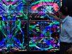
Professor Goebel: That is one of the main objectives. The current MRI scanners that work with magnetic field strengths of 1.5-tesla or 3-tesla have achieved a spatial resolution of two to three millimeters. In our Brain Imaging Center, we now also have two ultra-high field scanners with magnetic field strengths of 71 and 9.41 tesla. Simply put, you get a strong image signal with higher field strengths. You can zoom deep into the brain and measure activity in areas that are tiny. Only few have ever seen brain images like the ones we’re getting with our ultra-high field scanners.
Do you expect a qualitative leap in research?
Professor Goebel: Yes, we are profiting from a lucky fluke of nature. The cerebral cortex has characteristic functional units called the cortical columns.
These modules contain approximately 10,000 nerve cells that all do the same thing – for instance, they react the same way to a certain sensory input.
This represents a kind of redundancy principle in the brain. We are familiar with the column architecture, particularly in the perceptive areas of the cortex2, but there are probably comparable functional modules in most – if not all – areas of the brain. The main point is now that with the new ultra high-field scanners we may not be able to detect the activity of individual neurons, but we can detect individual functional modules since they are in a detectable range of the scanner. This means, for the first time, we might be able to improve our understanding of the code with which a sensory input is represented in the cortex.
Train your Brain
When around 150 internationally recognized experts in neuronal imaging met in Zurich, Switzerland in February 2012, it marked a milestone in the development of an emerging therapeutic discipline: brain-scan-based neuro feedback. Back then, the scientists discussed – at the first-ever international conference on the subject – the state and perspectives of the methods in which patients learn to control their brain activity.3
The principle involved is simple: While lying in a magnetic resonance imaging (MRI) scanner, the patient views a monitor that shows in real time how active a certain area of the brain is using, for example, an activity “thermometer.” Just as it is possible in other forms of bio feedback to control one’s heartbeat and blood pressure, the patient learns to decrease or increase brain activity in the respective region using specific mental strategies or fantasies – and can immediately see the success of their efforts in the scanner signals. A special software analyzes and visualizes the scanner data.
The idea of neuro feedback is not new. Neuro feedback based on electrical brain activity measurements (EEG) has been used for a long time, for example, to train people with epilepsy to suppress their seizures or to mitigate an Attention Deficit Hyperactivity Disorder (ADHD). However, the scanner-based process permits a deeper look into the brain and a much more precise analysis of activity patterns, which expands the spectrum of potentially treatable illnesses. Neuro feedback training is typically performed in several sessions in MRI machines with a field strength of 3-tesla.
The innovative approach has been developed over the past decade by a relatively small number of international specialists – including Rainer Goebel, who conducted basic research on scanner-based neuro feedback in close cooperation with German neuro researchers Niels Birbaumer und Nikolaus Weiskopf. Interest in the method has since grown considerably. Pilot studies by various teams show that self-regulation of brain activity can alleviate chronic pain, reduce motor deficits in Parkinson’s disease, or even improve rehabilitation after a stroke.4
The results in patients with depression are especially promising. A Phase I randomized clinical trial, which examines the benefits of scanner-based neuro feedback in regulating the emotional centers of the brain, is almost finished.5 There are more clinical trials on other diseases to follow as part of “BrainTrain,” an international collaborative research project supported by the European Commission.6

Can you give us an example?
Professor Goebel: Take facial recognition. What we already suspect from current experiments is that specific facial features – such as the distance between the eyes, the hairline or the position of the eyes in relation to the mouth – are coded by the activity of individual cortical columns. The brain breaks down (so to speak) the sensory input at the columnar level into different aspects, into a distributed code.
Based on a specific column pattern, it might be possible that the scanner has the ability to – in principle – discern whether you are looking at your partner’s face or the German Chancellor’s face. It makes a detailed reading of the brain activity possible.
Cortical column patterns probably also explain how the brain registers new faces and recognizes them again later.
What effects do you see for the field of medicine?
Professor Goebel: One example we’re currently working on is reading disabilities in children, also known as dyslexia. Thanks to the improved scanner resolutions, we hope to determine why and where letters are mixed up in the cortex through incorrectly coded column patterns and what kind of training can best influence this – in other words, how the brain’s plasticity can be used for treatment. But we also expect significant advances in our understanding of depression or schizophrenia.
Are there similar perspectives for neurodegenerative diseases such as Alzheimer’s?
Professor Goebel: With Alzheimer’s, we believe that it will at least be possible to make an impactful assessment – because the ultra-high field scanners will probably be able to detect signs of degeneration in the fiber connections of the brain. The high scanner resolution can also help with Parkinson’s when, for example, a patient is supposed to receive what’s called a “brain pacemaker” and precise neurosurgical planning is required for the surgery.
However, the advantages of imaging for treatment do not depend only on a maximum image resolution, but also on the possibility of having scanners that are integrated into new treatment concepts. For instance, for many years we have been examining the possibility of targeted neuro feedback with a 3-tesla scanner in which patients can learn to control the activity levels of individual areas in the brain using signals from the scanner (see info box entitled “Train your Brain“). This opens up completely new treatment paths for a wide range of illnesses, such as phobias or depression.
Do the modern images of the brain also change how we see ourselves?
Professor Goebel: We hope, in ten years, people may say: “Doctors determined in a brain scan that my left amygdala in the limbic system is overstimulated. That’s why I am going to neuro feedback therapy to be less afraid of spiders.” Overall, how mental processes work in detail may be much better understood and might even be included in textbooks. We may learn to talk much more concretely and realistically about our brain and ourselves. We will increasingly come to understand the mind in biological terms.
Are we losing our mystique due to fMRI?
Professor Goebel: No, because the images do not provide information about the subjective experience, the experience of our own consciousness. The I-perspective remains a mystery, even for us.
About the Author
Dr. Martin Lindner is an award-winning science writer based in Berlin, Germany. After his medical studies and a doctoral thesis in the history of medicine, he went into journalism. His writings have appeared in many major German and Swiss newspapers and magazines.