
History
A 67-year-old male patient, suffering from long-term pain in his left lower limb, presented himself to the hospital for a checkup. The pain commonly occurred after walking or an extended period in orthostasis. There was no associated local swelling. His medical history was unremarkable except for a long-term well-controlled hypertension. A Runoff CT angiography (CTA) was requested for evaluation.
Diagnosis
CTA images showed a dilated left femoral artery and vein, which along with the also dilated popliteal artery and vein, extended into a tangled and tortuous vascular mesh with a nidus. The anomalous vessels were accentuated posteriorly and distributed between the muscle bellies, with no signs of intra-articular or intraosseous extensions. Arteriovenous communications were evidenced, suggesting an arteriovenous malformation (AVM).
The patient underwent a vascular embolization and was in good general condition with improved symptomatic in the follow-up.
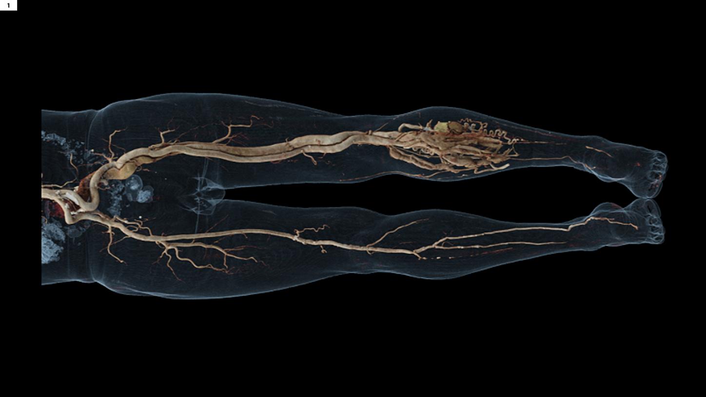
Fig. 1:
A cVRT image shows a vascular overview of the entire lower extremities in the arterial phase. The dilated left femoral artery and vein, as well as the popliteal artery and vein, extend into a tangled and tortuous vascular mesh with a nidus.
Comments
AVMs are vascular malformations composed of arteries and veins that directly communicate through a central nidus, bypassing the high resistance of capillary beds and leading to a high-flow lesion that shunts blood from arterial to venous circulation. Embolization alone, or in conjunction with surgical resection, is the primary treatment option for AVMs. Imaging evaluation is vital to the characterization and subsequent treatment planning. A Runoff CTA allows vascular assessment of the entire lower extremities, including the AVM and its relationship to the specific muscles and bones, in a single scan. In this case, image reconstruction of orthogonal planes, additionally to axial thin slices, was possible with the syngo.via platform, enabling detailed and fast vessel evaluation. Three-dimensional life-like image demonstration using cinematic volume rendering technique (cVRT) provides improved depth and shape perceptions.


Fig. 2:
Oblique MPR images show arteriovenous communications (arrows) evidencing an AVM.

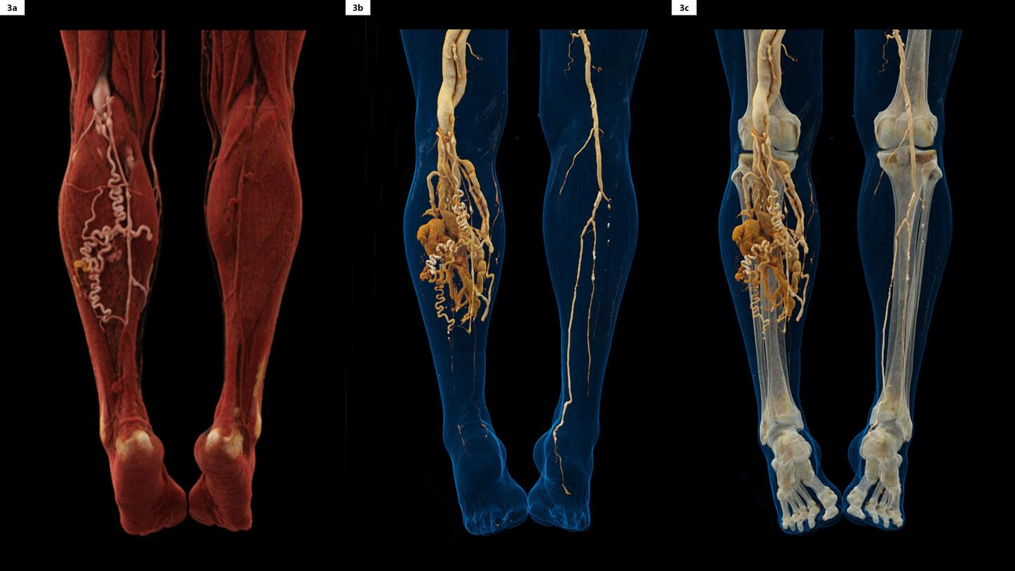


Fig. 3:
cVRT images, displayed in different presets, show the posterior view of the AVM in detail.