CT-guided interventionsInnovative CT solutions for efficient procedure planning and needle guidance
Our mission is to simplify and elevate your CT-guided interventions by delivering a modular, scalable solution tailored to meet your individual needs as a clinician.
We are dedicated to helping you achieve precise and efficient needle procedures through advanced path planning, AI‑driven needle detection, seamless 3D registration and image fusion, combined with laser-guided insertion.
With flexible in-room control options, we enable you to work confidently – whether independently or with assistance – ensuring smooth workflows and allowing you to focus entirely on providing the best possible patient care.
Highlights & Innovations
Explore our highlight technologies designed to facilitate rapid, precise, and secure routine as well as complex CT-guided procedures.

myNeedle Laser
Fully integrated laser guidance for even advanced procedures with multiple needle paths.
myAblation Guide
Comprehensive coverage from ablation planning to treatment assessment
Clinical applications
CT-guided interventions are expected to rise, with biopsies remaining common but therapeutic procedures growing faster due to benefits like fewer complications, faster recovery, and shorter hospital stays. Discover key clinical areas where CT-guided interventions are making a significant impact.
Biopsy
Biopsies remain essential for fast, reliable diagnoses. CT guidance improves needle placement accuracy and increases the success rate of obtaining diagnostic tissue during the initial procedure.1
Image courtesy of Hospital Maennedorf, Switzerland
Drainage
Drainage procedures are fundamental for quickly resolving critical fluid collections with minimal invasiveness. CT‑guided placement ensures safe, efficient workflows that help patients recover faster.2
Image courtesy of The Ohio State University Wexner Medical Center, USA
Pain management
Pain management is crucial as chronic conditions place a major burden on patients worldwide. CT‑guided injections deliver targeted relief with accuracy that reduces more invasive procedures.3
Image courtesy of Benson Mount Gambier Hospital SA, Australia
Tumor ablation
Pain management is crucial as chronic conditions place a major burden on patients worldwide. CT-guided injections of pain medication deliver targeted relief with accuracy that can help avoid more invasive procedures.4
Image courtesy of RNS Group Practice for Radiology and Radiotherapy Wiesbaden, Germany
Leverage our clinical applications for efficient procedure planning and needle guidance
To help you perform efficiently in each of these clinical fields, our CT‑guided solutions give you the clarity, control, and confidence you need throughout every step of an intervention. With intelligent planning tools, AI-based needle guidance, and flexible in‑room operation, you’re equipped to work smoothly and deliver consistently high‑quality care – even in complex procedures.
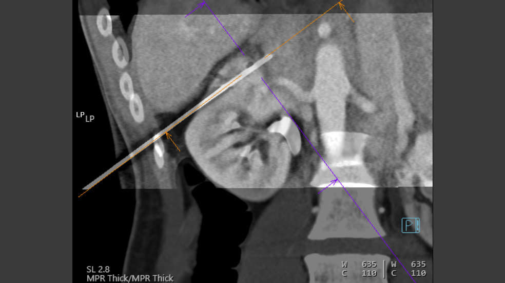
myNeedle Laser
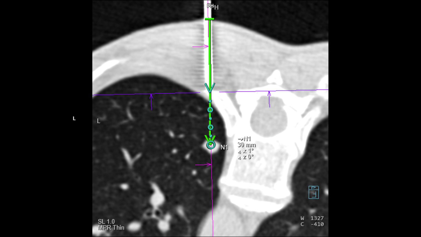
myNeedle Laser5 is a powerful, fully integrated option designed to make the intervention workflow more efficient by automatically projecting the needle entry point and insertion angle onto the body of the patient – even in advanced double-angulated procedures with multiple needle paths.
myAblation Guide
myAblation Guide6 brings unprecedented consistency to your microwave liver ablation workflow. With sophisticated planning tools, easy postprocedural assessment, and a coherent user experience across imaging and ablation systems, we’ve got you covered every step of the way.
myNeedle Guide 3D
myNeedle Guide 3D7 simplifies the workflow for both routine and complex CT-guided interventions. You can easily plan multiple needle pathways in different cross sections. Both path planning and visually guided insertion of multiple needles are supported within the workflow.
The AI based needle detection algorithm myNeedle Detection8 supports an efficient workflow by minimizing user interaction when progressing the needle.
Integrated Image Fusion allows fusing of 3D images from different modalities or contrast-enhanced prior CT studies to accurately plan and guide needle paths around critical anatomy.
Clinical cases

Courtesy of Kantonsspital Baden, Baden, Switzerland
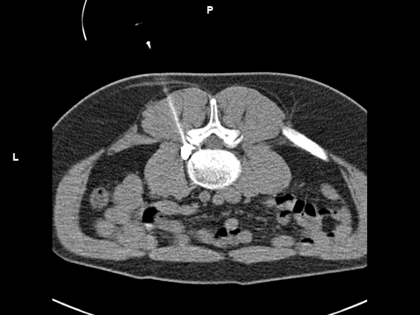
Biopsy of a small suspicious lesion at the shoulder blade
CT-guided intervention with myNeedle Guide 2D on SOMATOM X.ceed
Planning and control spiral:
Exposure time: 3.6 s / 1.7 s
Scan length: 131 mm / 41 mm
100 kV
CTDIvol: 3.9 mGy / 3.15 mGy
DLP: 57.6 mGy*cm / 18.7 mGy*cm
i-Fluoro:
3 events
130 kV
Scan length: 9 mm
Exposure time: 0.7 s / 4.3 s / 0.7 s
CTDIvol: 6.48 mGy / 39.3 mGy / 6.48 mGy
Accum. DLP: 47.1 mGy*cm
Complete procedure time: 36 min


myNeedle Guide 2D
myNeedle Guide 2D simplifies CT-guided interventions along the whole clinical workflow, from imaging through planning and monitoring the current needle position.
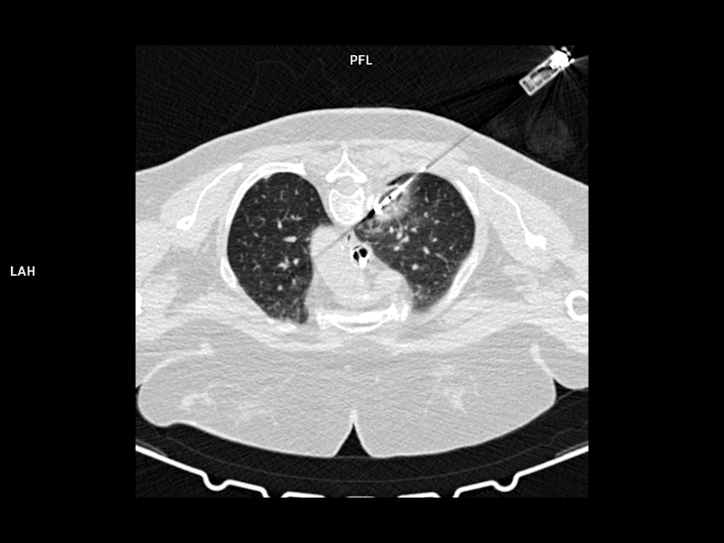
i-Fluoro
i-Fluoro9 mode is designed for precise and fast placement in even complex, moving anatomies. At the same time, it offers HandCARE a real-time dose reduction providing significant protection from radiation to both radiologist and patient.10
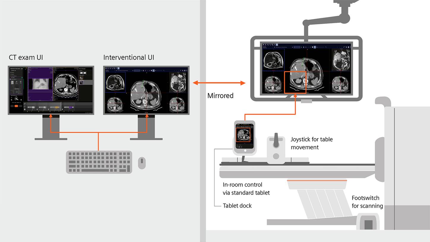
In-room control
Clinical practice
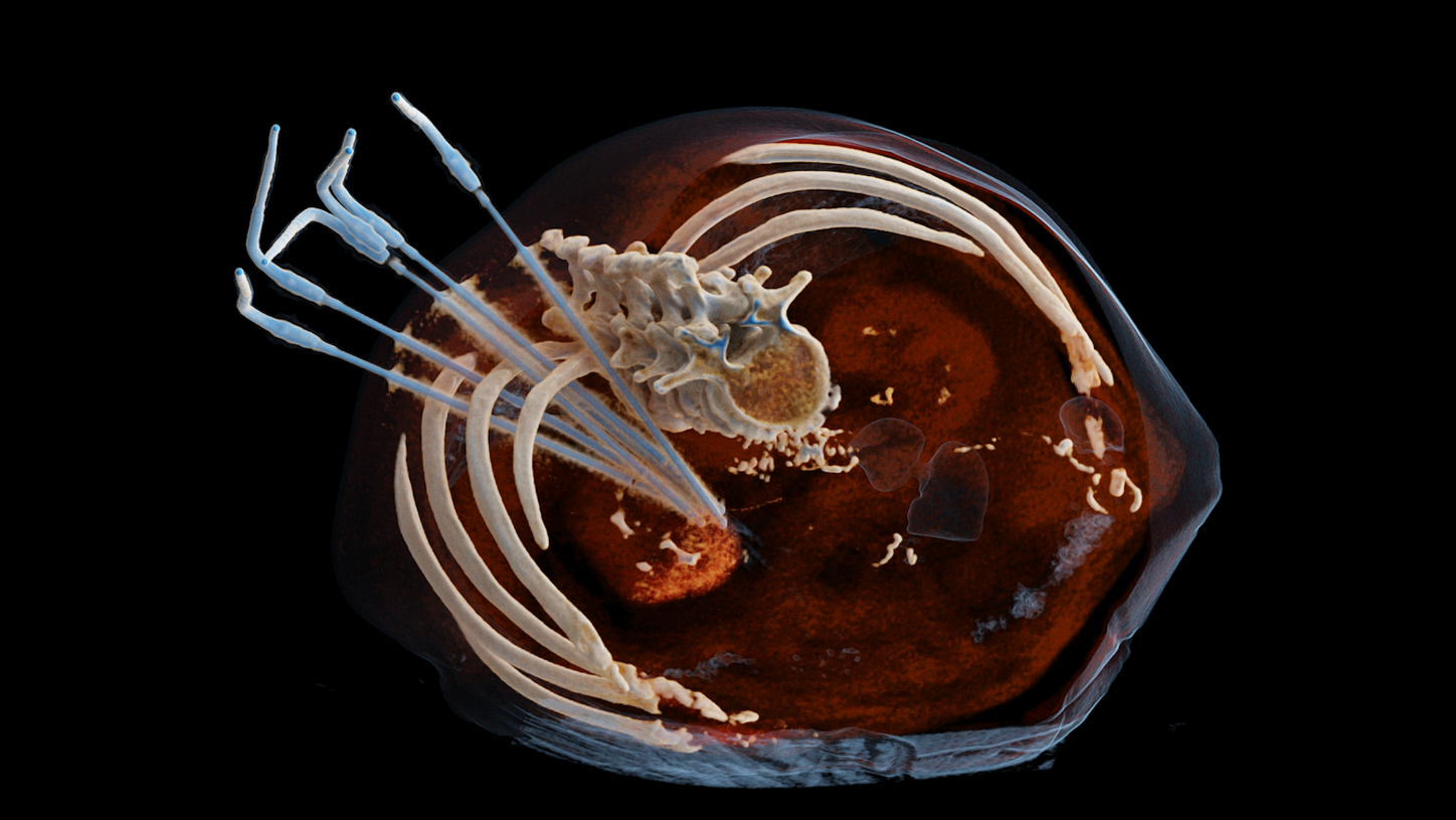
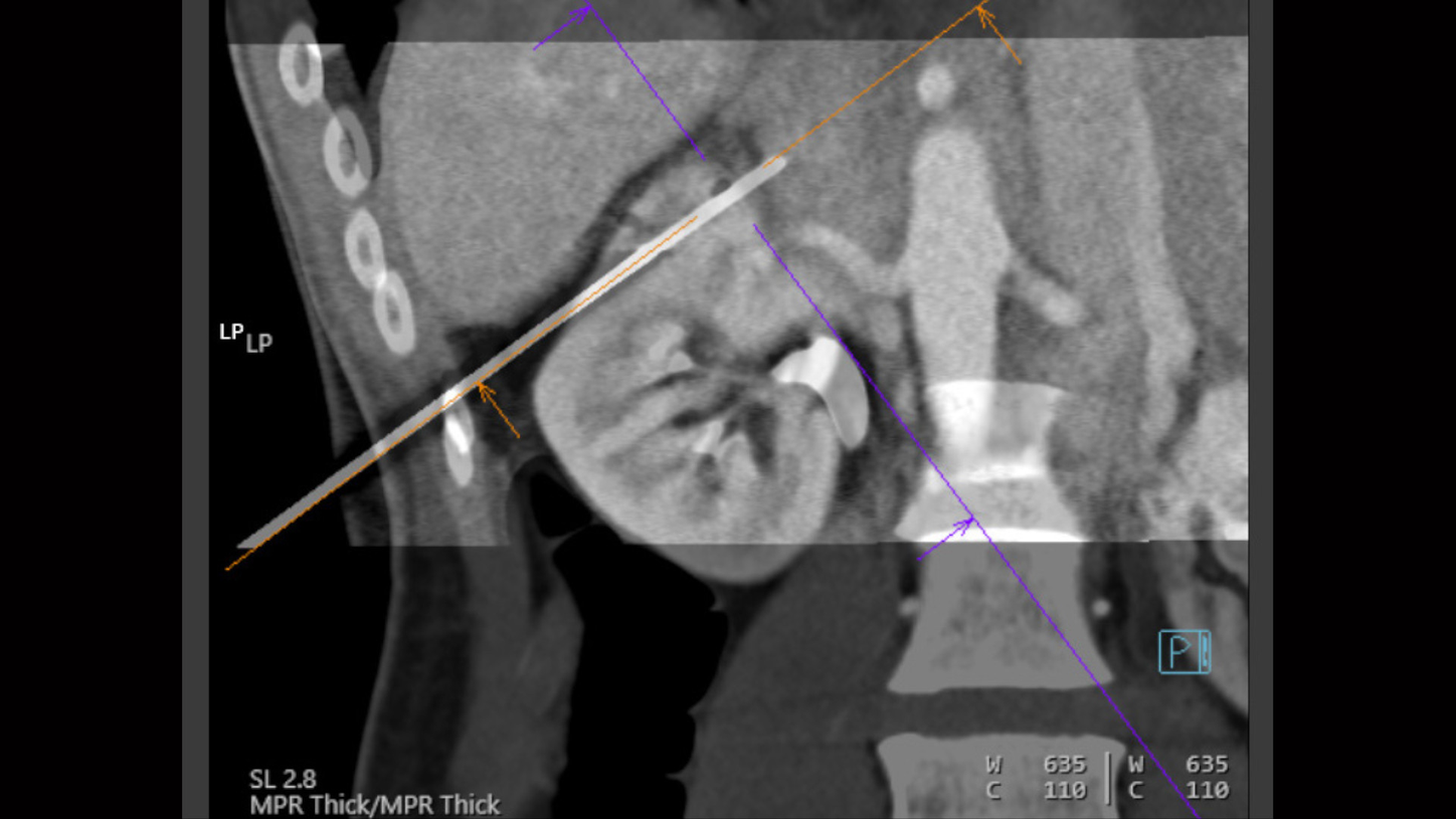
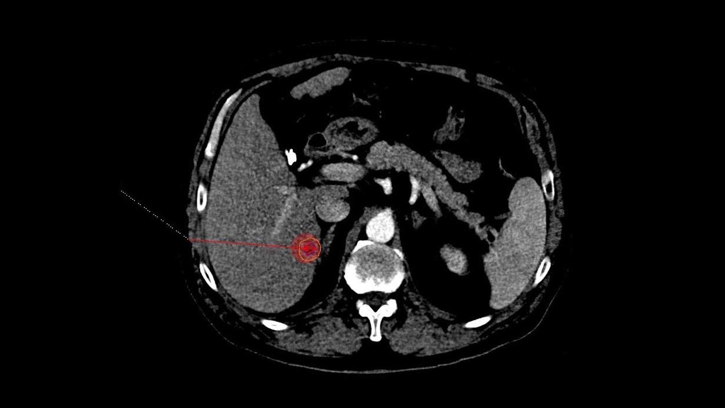
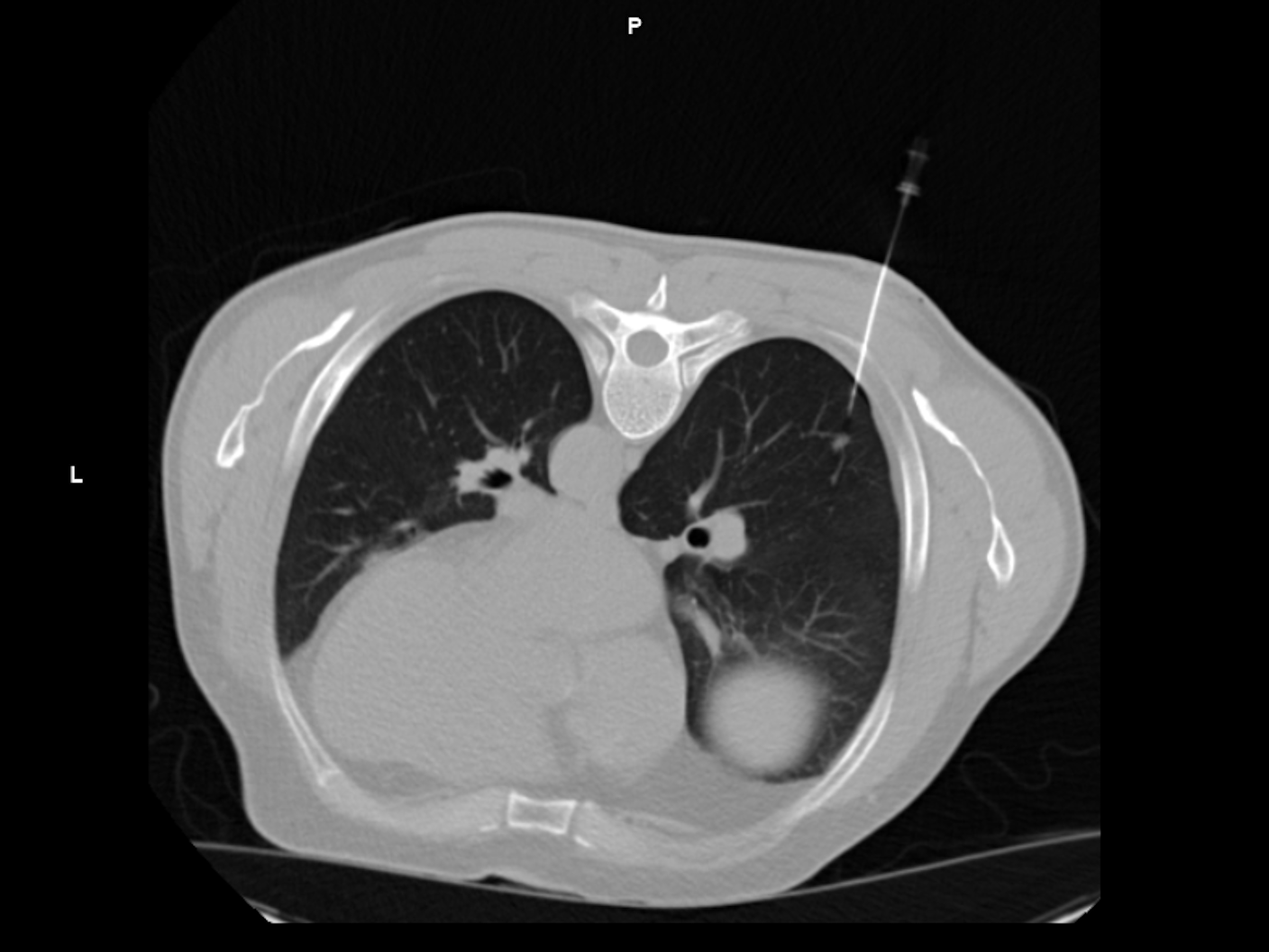
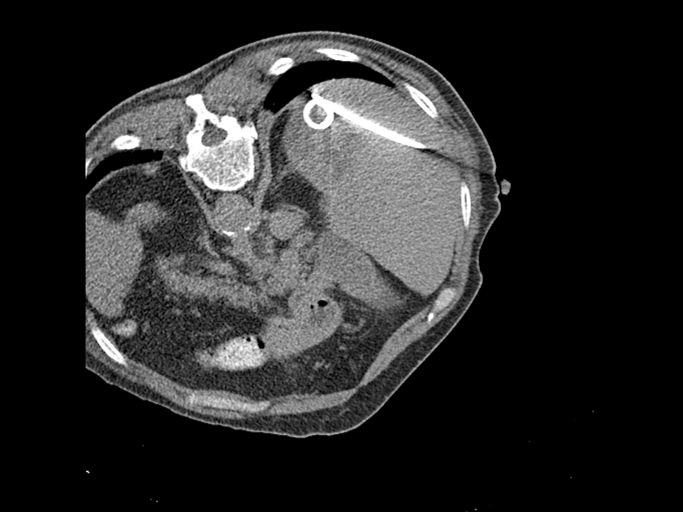
Cryoablation of an adrenal metastasis under CT guidance
Clinical Field: Interventional ablation in oncology
Products Used: SOMATOM X.ceed, myNeedle Guide 3D, myNeedle Laser
Case: An 85‑year‑old with a growing adrenal metastasis tightly located between the kidney and aorta required extremely precise probe placement, including a challenging transpulmonary approach.
Solution: Multi‑probe cryoablation planned via myNeedle Guide 3D for complex double‑angulated trajectories, transferred to myNeedle Laser for exact entry and angle visualization; progression monitored via i‑spiral CT.
Outcome: Six cryoprobes were placed accurately, no complications occurred, and follow‑up CT at 3 months showed complete devascularization with no recurrence.