Scimitar syndrome

Liguo Yao, MD1; Laihu Yang, MD1; Dalin Zhu, MD1; Jifang Qian, MD1; Pengyun Cheng, MD2; Xinglong Liu, MD2

1 Department of Radiology, Gansu Provincial Maternity and Child-care Hospital, Lanzhou, P. R. China

2 Siemens Healthineers, China

17.09.2021
A 6-year-old boy, suffering from a dry cough and fever for the past 10 days, was admitted to the hospital. Auscultation revealed bilateral rales and slightly suppressed respiration of the right lung. A CT chest examination was requested for evaluation.
CT images revealed a partial anomalous right pulmonary vein (PARPV) which drained into the upper part of the inferior vena cava (IVC) above the diaphragm. The right inferior pulmonary vein (RIPV), of small caliber, returned into the left atrium (LA). The right pulmonary artery (RPA) and the right lung appeared hypoplastic. The mediastinum was shifted to the right. The right upper and middle bronchi ended blindly, without arborization. No signs of cardiac abnormalities were seen.
A coronal MPR image and a cVRT image show a PARPV draining into the upper part of the IVC above the diaphragm. A RIPV, of small caliber, returns into the LA. The right lung is hypoplastic and the mediastinum is shifted to the right.
Courtesy of Department of Radiology, Gansu Provincial Maternity and Child-care Hospital, Lanzhou, P. R. China
Fig. 1: A coronal MPR image (Fig. 1a) and a cVRT image (Fig. 1b) show a PARPV draining into the upper part of the IVC above the diaphragm (arrows). A RIPV, of small caliber, returns into the LA. The right lung is hypoplastic and the mediastinum is shifted to the right.
A MIP image and a cVRT image show a hypoplastic RPA (arrows).
Courtesy of Department of Radiology, Gansu Provincial Maternity and Child-care Hospital, Lanzhou, P. R. China
Fig. 2: A MIP image (Fig. 2a) and a cVRT image (Fig. 2b) show a hypoplastic RPA (arrows).
A coronal MPR image show diverticular right upper and middle bronchi without arborization.
Courtesy of Department of Radiology, Gansu Provincial Maternity and Child-care Hospital, Lanzhou, P. R. China
Fig. 3: A coronal MPR image show diverticular right upper and middle bronchi (arrows) without arborization.

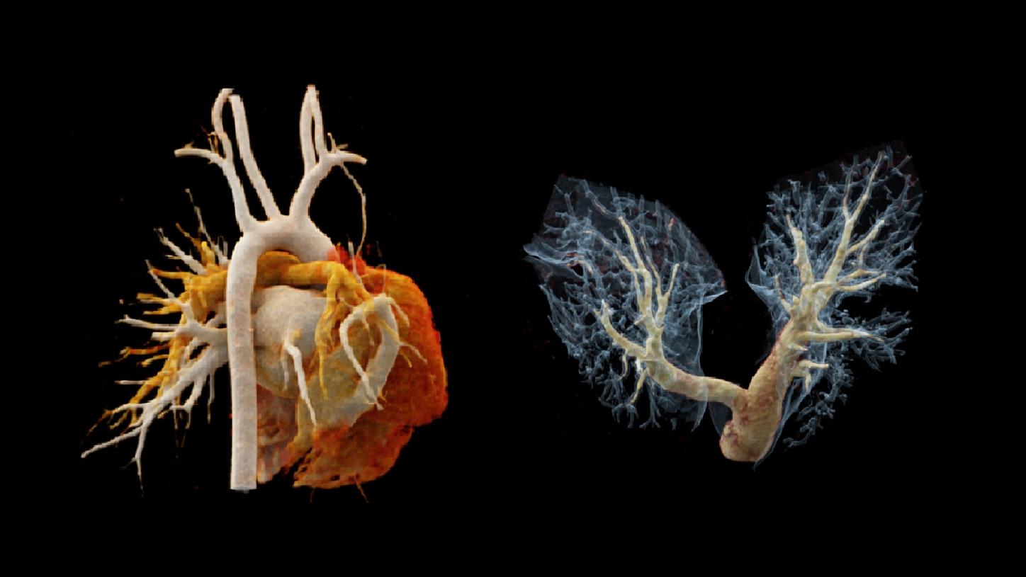
Scimitar syndrome is characterized by the combination of hypoplasia of the right lung and abnormal venous drainage to the inferior vena cava.[1] The syndrome comprises a wide spectrum of symptoms, making the diagnosis difficult, particularly in children and young adults with concomitant congenital cardiac and pulmonary anomalies. Surgical correction is usually carried out on symptomatic patients or on patients with an increased pulmonary blood flow and signs of right heart chamber dilation.[2] Imaging assessment prior to treatment is mandatory. In this case, a unique ultra-fast CT scan mode granted by dual source CT – “Turbo Flash mode” – is performed to complete a thorax scan in 0.42 s in free breathing. A lower kV setting, of 70 kV, is applied to enhance image contrast, optimizing image quality at a reasonably achievable lower radiation dose and less contrast agent. Three dimensional imaging techniques, such as multiplanar reconstruction (MPR), maximum intensity projection (MIP) and cinematic volume rendering technique (cVRT), offer the potential of higher diagnostic confidence and accuracy, as well as improving communication and planning for treatment.

Scanner

Scan area

Thorax

Scan mode

Turbo Flash mode

Scan length

209 mm

Scan direction

Cranio-caudal

Scan time

0.42 s

Tube voltage

70 / 70 kV

Effective mAs

154 mAs

Dose modulation

CARE Dose4D

CTDIvol

1.4 mGy

DLP

32.6 mGy*cm

Rotation time

0.25 s

Pitch

1.9

Slice collimation

192 x 0.6 mm

Slice width

0.75 mm

Reconstruction increment

0.5 mm

Reconstruction kernel

Bv40

Contrast

300 mg/mL

Volume

20 mL (70% contrast + 30% saline) + 10 mL saline

Flow rate

1.5 mL/s

Start delay

Bolus tracking with 100 HU at ascending aorta + 3 s