
Transrectal Ultrasound (TRUS)
- The gold standard for diagnosis of prostate cancer is the histological assessment of specimens obtained by TRUS-guided systematic core needle biopsy. The method involves taking tissue samples from the prostate in a geometric pattern.
- Advanced ultrasound options, such as color power Doppler and contrast-enhanced ultrasound, offer increased sensitivity and specificity for lesion detection and localization.
- Real-time elastography represents a further ultrasound technique that can help differentiate malignant from benign tissue for lesion detection and biopsy guidance.
Data courtesy of Nara Medical University Hospital, Nara, Japan
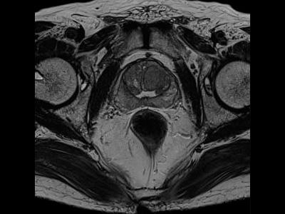
Magnetic Resonance Imaging (MRI)
- Multi-parametric MRI is used for tumor localization and delineation in a number of scenarios. Examples include negative TRUSguided biopsy with continued rise of PSA, relapse after initial therapy, local staging, and therapy planning.
- Siemens offers a full range of tumor staging and optimized prostate MRI applications such as syngo.MR General Engine Prostate Workflow & Report.
- A range of dedicated body, spine and endorectal coils are offered for the MAGNETOM 1.5 Tesla and 3 Tesla systems for optimized image quality.