SPECT/CT is a well-established imaging modality for investigation of suspicious bone lesions. But even so, additional testing is often required for confirmation of a patient’s situation—restricting everyone’s ability to deliver appropriate and timely care at a lower cost. The high image resolution of xSPECT Bone™ enables you to better localize and characterize disease, potentially reducing the need for alternative exams.
xSPECT Bone takes an entirely different approach in the generation of SPECT/CT images. By using the CT as the frame-of-reference for image reconstruction, xSPECT Bone is able to extract a zone map with different tissue segments to better delineate the boundaries of nuclear uptake during SPECT reconstruction. Through the deep integration of SPECT and CT data, xSPECT Bone can help you clearly distinguish disease not easily seen with conventional SPECT.
By boosting the level of clinically relevant information, xSPECT Bone imaging enables you to deliver much more specific and comprehensive reports to your referring physicians—facilitating better communication and more effective decision making.

xSPECT BoneEngineering a better bone scan
What experts say
" With xSPECT Bone, our reports have a higher level of information that can be acted on. This helps us better communicate with our referrers, so they can better manage their patients." 1

Insights
In August 2015, Garran Medical Imaging (GMI) in Canberra, Australia, initiated a study to prospectively evaluate their first 200 xSPECT Bone cases. As part of the study, GMI performed sequential reporting of SPECT/CT followed by xSPECT/CT. Differences between the initial SPECT/CT and the final report (after xSPECT/CT reconstruction) were documented and analyzed.
The findings, published in European Journal of Hybrid Imaging (March 2018), showed that xSPECT Bone imaging provided more diagnostic information in 71% of scans; and when compared to conventional SPECT/CT, the diagnosis was changed in 20% of cases.
Clinical image gallery


Case studies

xSPECT Bone used to delineate cervical vertebral screw loosening
By Iain Duncan, MD
Data courtesy of Garran Medical Imaging, Canberra, Australia
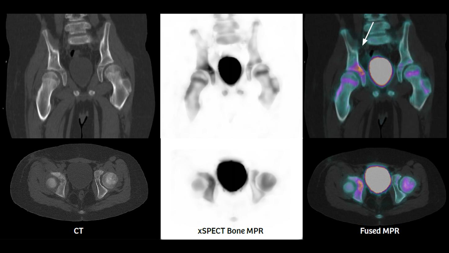
xSPECT Bone sharply delineates acetabular osteomyelitis in a 12-year-old boy
By Gary Marano, MD
Data courtesy of West Virginia University, Morgantown, West Virginia, USA
White paper

xSPECT Bone: a clinical overview
Partha Ghosh, MD
Siemens Healthineers
Molecular Imaging Business Line
Image comparison tool
See for yourself how xSPECT Bone can help improve diagnostic accuracy in the characterization of disease
xSPECT Bone on our Symbia Intevo SPECT/CT leverages the finite resolution and relevant clinical information from CT with the greater resolution and 3D iterative reconstruction of SPECT to create a single image that enables you to confidently deliver much more specific and comprehensive reports for bone scans to referring physicians.
Choose two image types and then use the slider options below to compare and see the difference for yourself:
- xSPECT Bone
- xSPECT Bone/CT
- CT
- 3D Iterative with AC
- 3D Iterative without AC