/

syngo.via pour IRM
/
Le secteur de la radiologie connaît des défis de plus en plus importants avec l’augmentation des cas quotidiens, de la pression sur les coûts et des attentes.
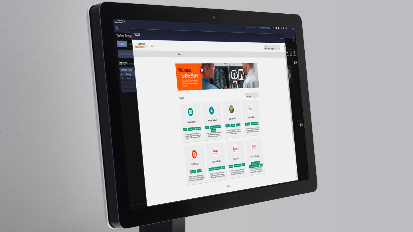
syngo.via est un logiciel intégré intelligent qui vous aide à affronter et surmonter ces défis. Grâce à une lecture multi-modalités et des résultats 3D rapides, vous pourrez avancer plus vite dans votre routine quotidienne. Le logiciel comporte les dernières innovations et fonctions recourant à l’IA afin de faire passer vos lectures au niveau supérieur. Il vous permet par ailleurs d’étendre et de personnaliser vos capacités de lecture et de rapport grâce à une plateforme ouverte.
- Les avantages des applications IRM dédiées pour des besoins spécifiques
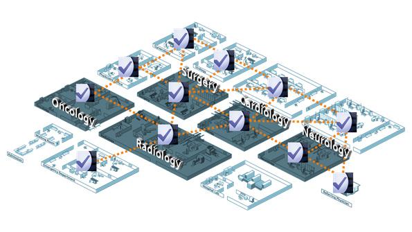
- Une véritable solution multi-modalités au service de tous les champs cliniques clés
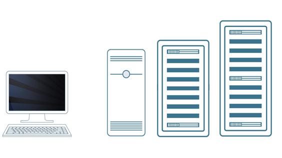
- Une flexibilité intelligente afin de répondre à vos exigences, aujourd’hui comme demain