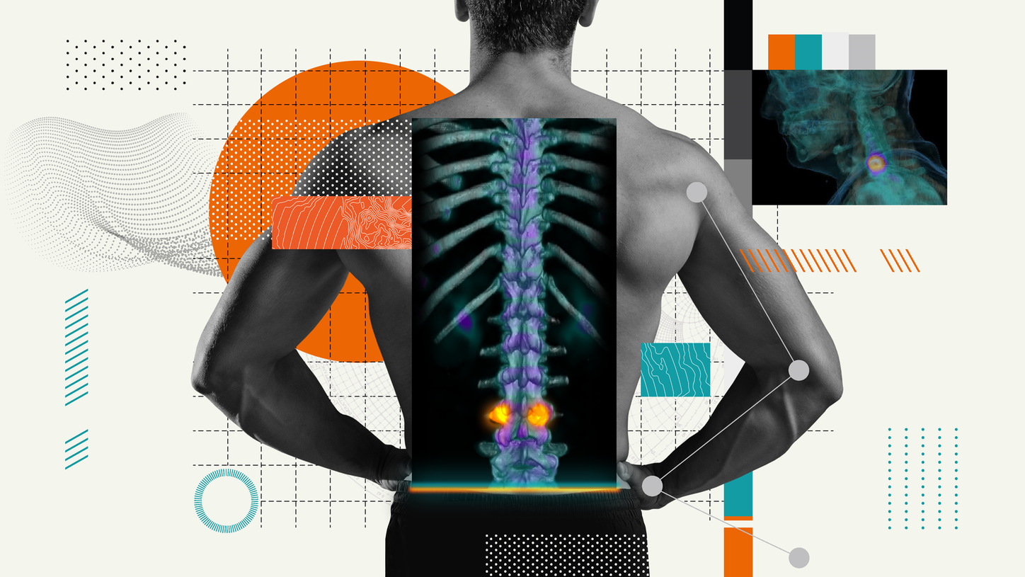
In 2009, Ethan Colliver, DO—a Physical Medicine and Rehabilitation physician—founded the Valley Sports and Spine Clinic in Virginia, USA, to treat back, shoulder, hip, and knee pain. Ten years later, Colliver finds that about 85% of the patients who come to his office in Blacksburg present with spinal pain; most have been referred by primary care providers and about half are aged 65 years or older. At the core of his practice, Colliver employs a wide range of image-guided, nonsurgical treatments for his patients. “X-rays, MRIs, and CT scans give us anatomic detail that can show whether there are potential pain generators. SPECT gives us physiological information, so it tells us if a particular area of degenerative arthritis (osteoarthritis) is physiologically active. The more degenerative a joint is, the higher chance we will see increased metabolic activity in that joint.” It is widely reported about 40% of the joints visible on SPECT fail to reveal severe arthritis on CT or other anatomic-based modalities, Colliver notes.
"When I view the images, whether I view increased metabolic activity from a degenerated joint or not, the image tells me something.”

Increased use of SPECT/CT
Colliver has utilized hybrid SPECT/CT imaging since 2014, although he was first introduced to it during his 2005-2008 residency at the University of Utah. “My mentor, Dr. Stuart Willick, was one of the pioneers in using SPECT/CT to localize axial spine pain,” Colliver recalls. “Dr. Willick called the technique a ‘fire scan‘ because areas of increased bone turnover lit up.1 He showed me a ‘fire scan’ of a young man who experienced back pain for months; just one facet joint was lit up, so Dr. Willick was able to do a single therapeutic injection in that joint.” But as Colliver moved on to other institutions, he found they were more skeptical about ‘fire scans’; he did not utilize SPECT/CT again until 2012, when he investigated its use for his own clinic. “Back then I was more likely to start with an MRI, and if I couldn’t figure out what was going on in an MRI, then I would do a SPECT/CT,” he says. “But over the years I have often found that if someone has back pain for a long time, I’ll go straight to SPECT/CT and avoid doing an MRI.” Nowadays Colliver orders SPECT/CT for 40-50% of his patients, most often for those with low-back pain. Scanning is performed by the imaging services department at Carilion Roanoke Memorial Hospital in Virginia, which Colliver acknowledges has been indispensable in his clinic’s ability to easily access SPECT/CT images and reports.

Diagnosing chronic back pain
“SPECT/CT is a great imaging modality that is underutilized for people with chronic back pain,” Colliver asserts. He orders SPECT/CT scans for almost all patients with chronic pain (present for at least 3-6 months) who primarily experience axial pain. “SPECT/CT is more likely to prove useful in older patients, so for patients aged 50 years and older, I’ll order one X-ray and then a SPECT/CT to help localize,” he says. Most back-pain patients are diagnosed with degenerative arthritis, but SPECT/CT can also identify fractures, infections, inflammatory autoimmune conditions, and cancer. SPECT/CT is also useful in determining the cause of trauma-related pain, such as in a young athlete who experiences back pain for a few months. In this case, SPECT/CT’s imaging capabilities can help determine whether the athlete has a pars fracture. “When I view the images, whether I view increased metabolic activity from a degenerated joint or not, the image tells me something,” Colliver stresses.

SPECT/CT-guided treatment
“When I find areas of severe uptake, if it is a fracture, I have to decide whether to treat with a brace or do kyphoplasty,” Colliver says. In cases of degenerative arthritis, it is important to determine whether the pain generator is from an anterior or posterior element of the spine. The majority of pain generators are located in either the anterior or the posterior spinal column, which determines the site for injection. “If there is arthritis in the anterior column, I will do an epidural steroid injection,” he notes. Radiofrequency ablation, a potentially longer-lasting treatment, is another option for the facet joints. “SPECT/CT can also help me decide whether I’m going to prescribe extension-based or flexion-based exercise,” he says. For an anterior-column problem, flexion will worsen the pain, whereas extension will lessen it. “For posterior-column degenerative arthritis, I focus on neutral mechanics and have the patient keep a neutral spine, avoiding extreme extension. If they have a disc herniation, they should do extension exercises that involve bending backwards.” Colliver believes more research is needed to determine the types of exercise that will benefit the different causes of back pain identified on SPECT/CT imaging. “Currently, all we know is that exercise is good for back pain.” People who exercise can decrease their risk of future pain episodes by about 45%.2 He further iterates that, “the next step is to confirm if we can treat appropriately based on what we learn.” His clinic is currently participating in a research study led by Jackson W. Kiser, MD, chief of molecular imaging at Carilion Roanoke Memorial Hospital, to evaluate the clinical impact of SPECT/CT on the management of patients with spinal pain.3
Healthcare benefits
Greater utilization of SPECT/CT could have a positive financial impact, Colliver believes, especially since back and neck pain currently represents the third-largest condition contributing to healthcare spending in the USA.4 “It’s a decrease in healthcare dollars because you need not order as much imaging. Then you are not doing as many procedures, the patient feels better faster, and the number of office visits should also decrease. I think it’s just a matter of getting the word out, so that people are familiar with when to use SPECT/CT,” Colliver advises.
About the author
Linda Brookes is a freelance medical writer and editor who divides her time between London and New York, working for a variety of clients in the healthcare and pharmaceutical fields.