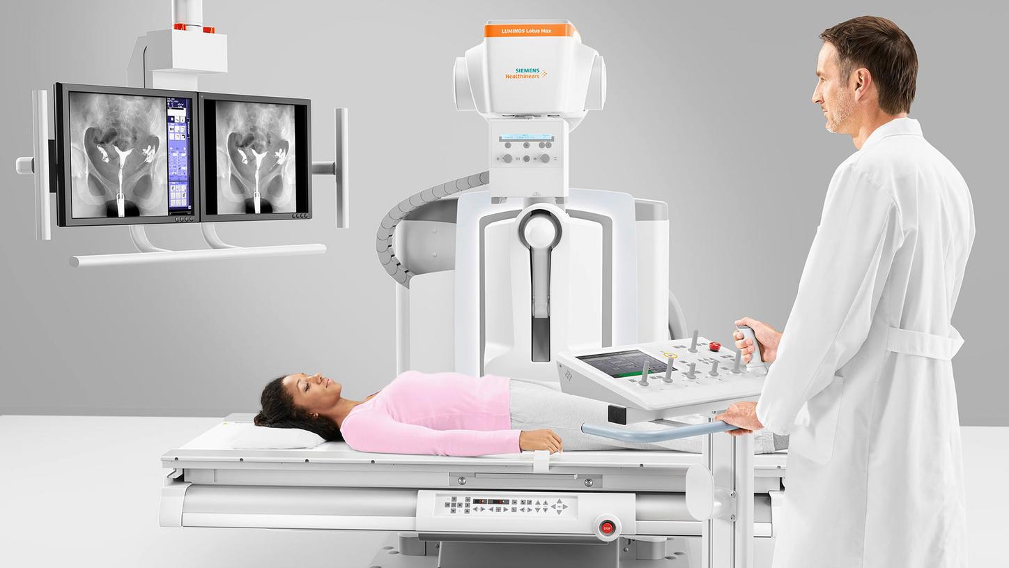
Pediatric patient undergoing procedure on LUMINOS Lotus Max with radiologist performing the clinical examination.

Rethinking dose in everyday imagingWhat impacts dose in radiography & fluoroscopy and where savings are possible

Dose outcomes are rarely driven by one factor alone. Instead, they reflect the interaction of technique, workflow, system design, and clinical environment.

Contributors to dose include:

Clinical Factors

  • Patient size & anatomy
  • Exam type & complexity
  • Area being imaged
  • Patient motion / positioning
  • Number of images / fluoroscopy time

Technical Factors

  • Exposure settings (kVp, mAs)
  • Beam collimation & filtration
  • Distance (source-to-patient/image)
  • Use of grid / detector sensitivity
  • Fluoroscopy settings (pulse rate, dose mode, magnification)

Operator/System Factors

  • Technique & protocol selection
  • Use of dose-reduction features
  • Equipment performance & QC
  • Repeat imaging

Dose reduction is possible

Dose reduction opportunities often exist through optimization of imaging parameters and examination protocols. Adjusting system settings while maintaining clinical objectives may help reduce radiation exposure for patients and staff.

Why this matters now

While median occupational doses for many imaging professionals remain relatively low, variability persists. Higher exposure is consistently associated with close patient proximity, high procedure frequency, and complex fluoroscopic workflows. As case volumes and procedural complexity increase, dose awareness becomes an operational issue—not just a physics consideration.

This makes dose optimization less about limits and more about designing imaging environments that support good decisions by default.

Subscription Button Icon
Be the first to know about our events, training, and news