Cardiac MR (CMR) exams in under 30 minutes—what once seemed impossible is now a reality. At Siemens Healthineers, we make it possible through highly automated, free-breathing exams that extend cardiac MR to a broader range of patients, delivering accessible, comfortable, and fast exams without compromise. Find out how below.

Cardiovascular MRITransform your CMR practice with cutting-edge applications
Highlights

3D WholeHeart Pro
3D WholeHeart Pro addresses complex cardiovascular MR imaging needs, offering high isotropic resolution and 3D tissue characterization essential for evaluating small lesions and extensive tissue damage.
The technique incorporates advanced automation algorithms for virtually artifact-free images and accurate coronary artery visualization in free-breathing for patients who struggle with breath-holds. 3D WholeHeart Pro integrates AI-based automation tools like AutoPositioning, AutoRestingPhase, and AutoTI streamlining the imaging process.
Cardiovascular applications
Free breathing exams
3D WholeHeart Pro
3D WholeHeart Pro addresses complex cardiovascular MR imaging needs, offering high isotropic resolution and 3D tissue characterization essential for evaluating small lesions and extensive tissue damage.
The technique incorporates advanced automation algorithms for virtually artifact-free images and accurate coronary artery visualization in free-breathing for patients who struggle with breath-holds. 3D WholeHeart Pro integrates AI-based automation tools like AutoPositioning, AutoRestingPhase, and AutoTI streamlining the imaging process.
Compressed Sensing Cardiac Cine
The Compressed Sensing segmented technique supports high spatial and temporal resolution with very short breath-holds. The Compressed Sensing real-time technique enables high-quality diagnostic imaging in free-breathing. These techniques can dramatically speed up cardiac scans, increase efficiency and expand the patient population eligible for Cardiac MRI, by supporting those with arrythmias or inability to hold their breath.

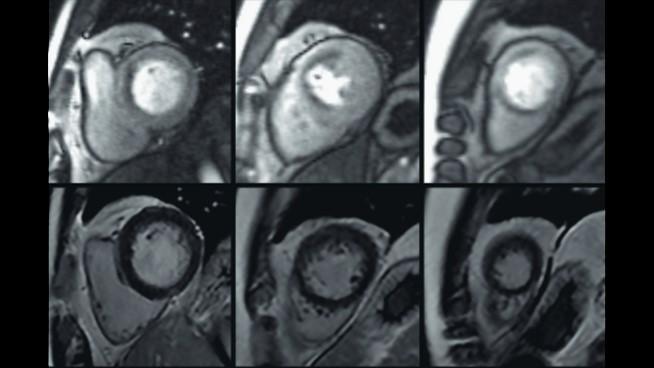
PSIR HeartFreeze
PSIR HeartFreeze with motion compensation algorithms enables high-resolution imaging in free-breathing for viability assessment, even in patients with arrhythmias and those who cannot hold their breath.
Highly efficient, automated CMR exams
Dedicated CMR appliactions
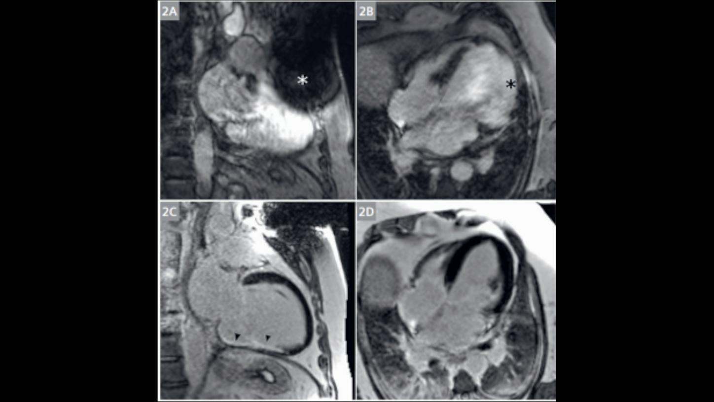
High Bandwidth Inversion Recovery
High Bandwidth Inversion Recovery (HBIR) significantly improves the quality of CMR imaging in patients prone to susceptivity artefacts. This advanced sequence employs wideband techniques to minimize distortions, which enhances tissue characterization for optimal imaging of myocardial injuries.
HBIR is particularly valuable as it extends high-quality diagnostic capabilities to patients prone to metal artefacts and thereby expands the potential for accurate disease management and diagnosis.

MyoMaps
MyoMaps facilitates pixel-based quantification of myocardial tissue, aiding in the differential diagnosis of myocardial diseases. When used in conjunction with HeartFreeze, our motion correction algorithm, MyoMaps enables the inline generation of T1, T2, and T2* colored parametric maps.

4D Flow
Flow quantification provides crucial insights into cardiac hemodynamics and function. ECG-triggered 2D phase contrast imaging enables non-invasive, quantitative assessment of blood flow. With retrospective reconstruction, full heart cycle coverage is achieved, allowing comprehensive analysis.
Our enhanced 4D Flow acquisition offers complete volumetric coverage, significantly improving the orientation and visualization of blood flow and velocity throughout the heart and thorax. This technical advancement facilitates the detailed analysis of key hemodynamic parameters over the entire cardiac cycle.
MR Angiography

QISS
QISS technology provides an accurate, non-contrast alternative for visualizing arterial disease, especially in patients where contrast agents are contraindicated or those with renal dysfunction.
QISS MRA is an easy-to-use approach for imaging peripheral arteries and contributes to patient safety by reducing the need for contrast agents and the associated risks.
Our dedicated MRI Scanner

MAGNETOM Sola Cardiovascular Edition
Outcome relevant decisions – redefining patient pathways
MAGNETOM Sola Cardiovascular Edition automatically adjusts to patient biovariability to overcome variations in cardiac MRI examinations:
- Free breathing exams
- Tissue characterization with MyoMaps and HeartFreeze
- CMR for challenging patients with High Bandwidth Inversion Recovery
- Highly efficient CMR exams with BioMatrix Beat Sensor and our automated exam assist tools
Clinical gallery
/


/