
Dual Source CT in functional imaging
Explore untapped potential
CT departments have employed large coverage 4D imaging and Dual Energy spectral imaging, both done at low radiation dose with Dual Source CT to advance the possibilities of functional imaging. Watch the videos below for examples of how you can continue to push the boundaries with Dual Source CT.
Explore untapped potential in functional imaging now
Visualization of hepatic lesions with CT perfusion
Earlier detection of hepatic lesions is critical for better patient outcomes. However routine CT and MR imaging are not sensitive enough. With Dual Source CT, take advantage of large-coverage low-dose perfusion protocols to get hemodynamic parameters to help detect these lesions and predict early response to anti-cancer treatment.
Visualization of pancreatic lesions with CT perfusion
Standard contrast-enhanced biphasic CT scans may fail to detect pancreatic lesions. Dual Source CT and its large-coverage CT perfusion protocols offer the possibility to get qualitative and quantitative information such as blood flow and blood volume that can help to visualize insulinomas - a special pancreatic tumor type.
Assessment of bone marrow edema with Dual Source Dual Energy
Employ Dual Source Dual Energy CT for assessment of bone marrow edema. Benefit from high sensitivity and high diagnostic accuracy for the assessment of subchondral insufficiency fracture (SIF) associated bone marrow edema. And potentially use it as an alternative to MRI for patients with contraindications or in situations where MRI is not available.
Evaluation of chronic thromboembolic pulmonary hypertension (CTEPH) with Dual Source Dual Energy
CTEPH has a poor prognosis if not treated and is difficult to diagnose. With Dual Source Dual Energy CT, you can get morphologic and functional pulmonary assessment without the need to use radioisotopes for scintigraphy - and benefit from high spatial resolution.
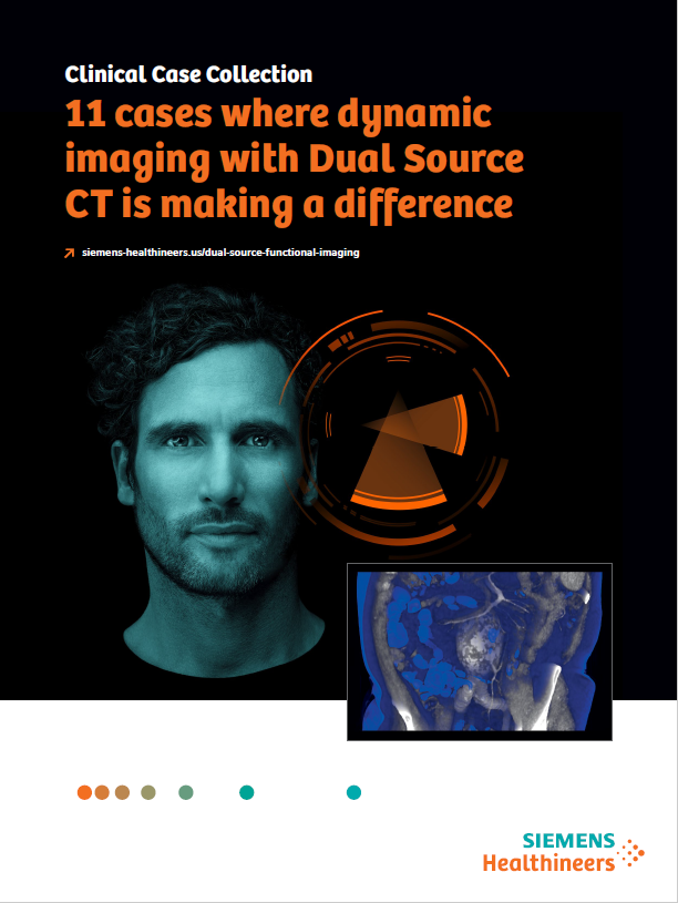
Case Collection: Where dynamic imaging with DSCT is making a difference
Morphological imaging alone may not be sufficient for advanced clinical cases like identification of vascular malformations or visualization of hepatocellular carcinoma or pancreatic lesions.
Dynamic imaging on Dual Source CT can make a difference – providing hemodynamic information at high dose efficiency and enough sensitivity to detect micro metastases and to demonstrate the feeding artery. Find 11 examples in a collection of clinical cases that demonstrate the power of Dual Source CT.
On-demand Webinar: Dual Source – Dual Energy CT in Clinical Practice
Bari Dane, MD, reviews common uses, benefits, and implementation of Dual Source – Dual Energy CT in clinical practice, as well as the benefits of a 3D camera used in conjunction with a Dual Source – Dual Energy CT scanner.