- Home
- Medical Imaging
- Computed Tomography
- SOMATOM
- SOMATOM Drive

SOMATOM Drive
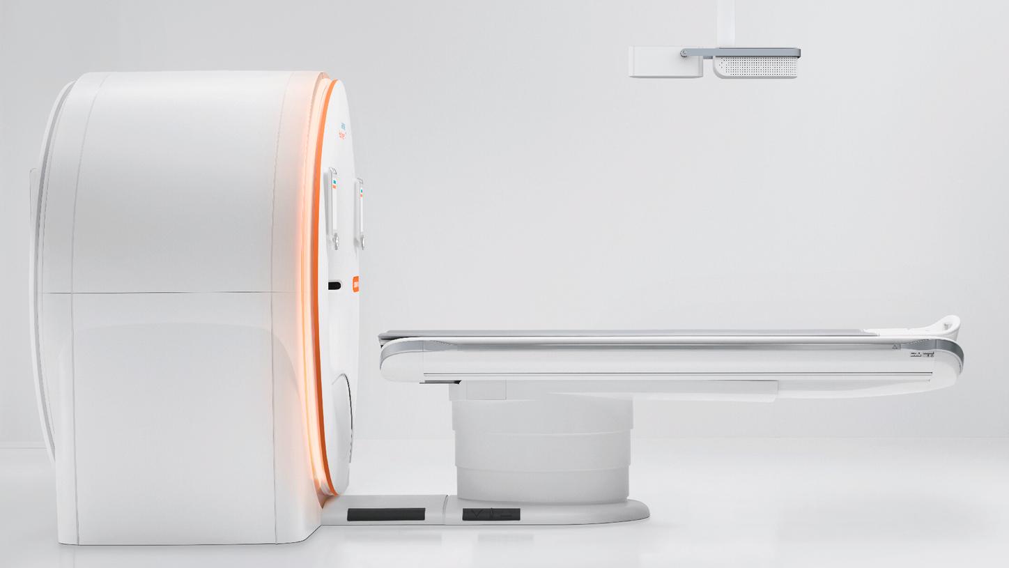
Drive precision for all – with a Dual Source CT scanner
Patients of all ages, sizes, and conditions arrive in your facility daily. And, you will be measured by how effectively you can care for each of them. They—and their referring physicians—need fast, clinically accurate information. In CT imaging, you can now reach a higher level of clinical confidence in your images across these many different case types while still meeting your efficiency goals. SOMATOM Drive with Dual Source technology boosts your performance, empowers your routines, and expands your capabilities.
Features & Benefits


Drive precision in bariatrics

Drive precision for your patients

Drive precision for pediatrics

Drive precision for urgent care

Drive precision for critical care

Drive precision for lung imaging

Drive precision in cardiovascular

Drive precision in orthopedics

Drive precision in bariatrics

Drive precision for your patients

Drive precision for pediatrics

Drive precision for urgent care

Drive precision for critical care

Drive precision for lung imaging

Drive precision in cardiovascular

Drive precision in orthopedics

Drive precision in bariatrics
Drive precision for your environment
SOMATOM Drive supports you in simplifying routines – to optimize performance and accelerate workflows. See how you can easily standardize your quality of care with our single-operator concept.

Safeguard correct and consistent patient positioning
FAST Integrated Workflow is a combination of unique solutions – such as the FAST 3D Camera, Touch Panels, and Advanced Applications – allowing:
- highly accurate patient positioning
- consistent image quality and dose
- improved workflow speed
- staff to stay close to their patients
Automation drives precision in challenging environments
Support precise patient positioning regardless of who is performing the scan, with full and semi-automated FAST and CARE features.
Automation drives precision in overburdened environments
Advance reading capabilities with reduction of metal artifacts (iMAR) and automatically aligned and labeled spinal images (FAST Spine).
Drive precision for your business needs
Our CT scanner is versatile and future ready: The Dual Source technology in SOMATOM Drive may advance new clinical fields and introduce new technologies into clinical practice.

Be ready for new business opportunities
Boost the reputation of your clinical institution with both patients and referrers with Dual Energy, dynamic imaging, and full organ perfusion.
Clinical Use
Cardiac Imaging
07Head and Neck Imaging
02Head and Neck Imaging
Thoraco-Abdominal Imaging
02Thoraco-Abdomen Imaging
01Pediatric Imaging
01
70 kV
Scan time: 0.33 s
Scan length: 142 mm
CTDIvol: 0.54 mGy
DLP: 11.3 mGy*cm
Eff. dose: 0.82 mSv
- Pediatric thoracic CT with free breathing and no sedation, patient rolling at time of scan
- 70 kV – CARE Child protocols can be performed with Turbo Flash scan modes
- Lower dose in highly sensitive patients
- Scan even a moving patient, as seen in this case
Courtesy of Medical University of Vienna, General Hospital AKH, Vienna, Austria

80 kV
Scan time: 0.29 s
Scan length: 135 mm
CTDIvol: 1.36 mGy
DLP: 25.4 mGy*cm
Eff. dose: 0.36 mSv
HR: 47-70 bpm (unstable)
- Coronary artery scan for assessment of vessel disease
- Outstanding image quality utilizing Turbo Flash mode (one heartbeat)
- Precise patient dose selection with High Power 80
Courtesy of Medical University of Vienna, General Hospital AKH, Vienna, Austria

80 kV
Scan time: 0.20 s
Scan length: 96.9 mm
CTDIvol: 1.42 mGy
DLP: 13.75 mGy*cm
Eff. dose: 0.19 mSv
HR: 60 bpm
- Coronary artery disease assessment with multiple calcified plaques
- High Power and 10 kV Steps enable precise patient dose selection
- Turbo Flash mode for low- dose, low-motion imaging
Courtesy of Amakusa Medical Center, Kumamoto, Japan

80 kV
Scan time: 0.21 s
Scan length: 101.8 mm
CTDIvol: 1.47 mGy
DLP: 14.9 mGy*cm
Eff. dose: 0.2 mSv
HR: 61 bpm
- Coronary artery scan for assessment of CAD
- High Power and 10 kV Steps enable precise patient dose selection
- Turbo Flash mode for low-dose, low-motion imaging
Courtesy of Amakusa Medical Center, Kumamoto, Japan

120 kV
Scan time: 0.73 s
Scan length: 322.5 mm
CTDIvol: 5.43 mGy
DLP: 175.1 mGy*cm
Eff. dose: 2.45 mSv
HR: <65 bpm
- Long-range coronary bypass follow-up scan
- Precise patient dose even with full functional study incl. valves & LIMA bypass graft at below 7 mSv
- Long-range cardiac imaging without compromising image quality
Courtesy of Amakusa Medical Center, Kumamoto, Japan

100 kV
Scan length: 143 mm
CTDIvol: 50.55 mGy
DLP: 721.0 mGy*cm
Eff. dose: 10 mSv
HR: 50 (49-53)
5 min delay (Dual Energy)
100/Sn140 kV
Scan length: 131 mm
CTDIvol: 52.57 mGy
DLP: 689.5 mGy*cm
Eff. dose: 9.6 mSv
HR: 60 (58-61)
- Post-stent follow-up with delayed enhancement visualizing myocardial scar
- Dual Energy with high temporal resolution for precise myocardial spectral imaging
- High-resolution stent imaging due to StellarInfinity detector
Courtesy of Amakusa Medical Center, Kumamoto, Japan
80 kV
Scan time: 0.54 s
Scan length: 282 mm
CTDIvol: 0.97 mGy
DLP: 27.4 mGy*cm
Eff. dose: 0.41 mSv
HR: 50 bpm
- Gated heart and aorta scan for work-up of ascending aortic aneurysm
- Turbo Flash mode with ECG gating
- Scan range beyond detector width in one heartbeat
Courtesy of Northwestern Memorial, Chicago IL, USA

110 kV
Scan time: 0.57 s
Scan length: 263 mm
CTDIvol: 3.87 mGy
DLP: 101.7 mGy*cm
Eff. dose: 1.42 mSv
HR: 61 bpm
- Gated heart and aorta scan for coarctation assessment
- Turbo Flash mode with ECG gating
- Scan range beyond detector width in one heartbeat
- 10 kV Steps for precise dose selection
Courtesy of Northwestern Memorial, Chicago IL, USA

80/Sn140 kV
Scan time: 24.2 s
Scan length: 160 mm
CTDIvol: 39.17 mGy
DLP: 626.7 mGy*cm
Eff. dose: 1.3 mSv
- CT brain for assessment of cerebral bleeding
- Clinically routine brain imaging with Dual Energy
- Enabled by wide spectral separation and dose-neutral DSDE
Courtesy of Amakusa Medical Center, Kumamoto, Japan

Sn100 kV
Scan time: 12 s
Scan length: 105.6 mm
CTDIvol: 1.65 mGy
DLP: 20.9 mGy*cm
Eff. dose: 0.04 mSv
- Spectral-shaped (Sn100 kV) sinusitis examination
- Tin-filtered scans reducing sinus CT dose to X-ray levels
Courtesy of Medical University of Vienna, General Hospital AKH, Vienna, Austria

80 kV
Scan time: 8.47 s
Scan length: 585.2 mm
CTDIvol: 3.63 mGy
DLP: 315 mGy*cm
Eff. dose: 3.9 mSv
- Full-body angiography for severe aortic dissection work-up
- High Power 80 driving low- dose, low-kV imaging, driving precision for all
Courtesy of Medical University of Vienna, General Hospital AKH, Vienna, Austria

Sn100 kV
Scan time: 0.72 s
Scan length: 330 mm
CTDIvol: 0.43 mGy
DLP: 14.2 mGy*cm
Eff. dose: 0.4 mSv
- Patient with shortness of breath for lung assessment
- Ultra-low-dose scan with Tin Filter and Turbo Flash mode
Courtesy of Medical University of Vienna, General Hospital AKH, Vienna, Austria

90 kV
Scan time: 4.53 s
Scan length: 448 mm
CTDIvol: 3.68 mGy
DLP: 165 mGy*cm
Eff. dose: 2.4 mSv
- Abdominal aortic scan for assessment of vascular pathology
- High Power and 10 kV Steps enable precise patient dose selection even in larger patients
Courtesy of Spire Manchester, Manchester, United Kingdom

100/Sn140 kV
Scan time: 14.8 s
Scan length: 342 mm
CTDIvol: 4.84 mGy
DLP: 165.5 mGy*cm
Eff. dose: 2.4 mSv
- Active bleeding indicating new fracture
- Clinically routine bone marrow imaging with Dual Energy
- Enabled by wide spectral separation and dose-neutral DSDE
Courtesy of Amakusa Medical Center, Kumamoto, Japan

70 kV
Scan time: 0.33 s
Scan length: 134 mm
CTDIvol: 0.28 mGy
DLP: 5.6 mGy*cm
Eff. dose: 0.52 mSv
- Thoracic CT without breath-hold – patient with congenital malformations of the thoracic vessels
- Turbo Flash mode scans at 75 ms temporal resolution enabling high image quality at low dose
- High power 70 enables low dose and contrast media (volume and concentration)
Courtesy of Erasmus MC, University Medical Center Rotterdam, Rotterdam, The Netherlands

70 kV
Scan time: 0.33 s
Scan length: 142 mm
CTDIvol: 0.54 mGy
DLP: 11.3 mGy*cm
Eff. dose: 0.82 mSv
- Pediatric thoracic CT with free breathing and no sedation, patient rolling at time of scan
- 70 kV – CARE Child protocols can be performed with Turbo Flash scan modes
- Lower dose in highly sensitive patients
- Scan even a moving patient, as seen in this case
Courtesy of Medical University of Vienna, General Hospital AKH, Vienna, Austria

80 kV
Scan time: 0.29 s
Scan length: 135 mm
CTDIvol: 1.36 mGy
DLP: 25.4 mGy*cm
Eff. dose: 0.36 mSv
HR: 47-70 bpm (unstable)
- Coronary artery scan for assessment of vessel disease
- Outstanding image quality utilizing Turbo Flash mode (one heartbeat)
- Precise patient dose selection with High Power 80
Courtesy of Medical University of Vienna, General Hospital AKH, Vienna, Austria

80 kV
Scan time: 0.20 s
Scan length: 96.9 mm
CTDIvol: 1.42 mGy
DLP: 13.75 mGy*cm
Eff. dose: 0.19 mSv
HR: 60 bpm
- Coronary artery disease assessment with multiple calcified plaques
- High Power and 10 kV Steps enable precise patient dose selection
- Turbo Flash mode for low- dose, low-motion imaging
Courtesy of Amakusa Medical Center, Kumamoto, Japan

80 kV
Scan time: 0.21 s
Scan length: 101.8 mm
CTDIvol: 1.47 mGy
DLP: 14.9 mGy*cm
Eff. dose: 0.2 mSv
HR: 61 bpm
- Coronary artery scan for assessment of CAD
- High Power and 10 kV Steps enable precise patient dose selection
- Turbo Flash mode for low-dose, low-motion imaging
Courtesy of Amakusa Medical Center, Kumamoto, Japan

120 kV
Scan time: 0.73 s
Scan length: 322.5 mm
CTDIvol: 5.43 mGy
DLP: 175.1 mGy*cm
Eff. dose: 2.45 mSv
HR: <65 bpm
- Long-range coronary bypass follow-up scan
- Precise patient dose even with full functional study incl. valves & LIMA bypass graft at below 7 mSv
- Long-range cardiac imaging without compromising image quality
Courtesy of Amakusa Medical Center, Kumamoto, Japan

100 kV
Scan length: 143 mm
CTDIvol: 50.55 mGy
DLP: 721.0 mGy*cm
Eff. dose: 10 mSv
HR: 50 (49-53)
5 min delay (Dual Energy)
100/Sn140 kV
Scan length: 131 mm
CTDIvol: 52.57 mGy
DLP: 689.5 mGy*cm
Eff. dose: 9.6 mSv
HR: 60 (58-61)
- Post-stent follow-up with delayed enhancement visualizing myocardial scar
- Dual Energy with high temporal resolution for precise myocardial spectral imaging
- High-resolution stent imaging due to StellarInfinity detector
Courtesy of Amakusa Medical Center, Kumamoto, Japan
80 kV
Scan time: 0.54 s
Scan length: 282 mm
CTDIvol: 0.97 mGy
DLP: 27.4 mGy*cm
Eff. dose: 0.41 mSv
HR: 50 bpm
- Gated heart and aorta scan for work-up of ascending aortic aneurysm
- Turbo Flash mode with ECG gating
- Scan range beyond detector width in one heartbeat
Courtesy of Northwestern Memorial, Chicago IL, USA

110 kV
Scan time: 0.57 s
Scan length: 263 mm
CTDIvol: 3.87 mGy
DLP: 101.7 mGy*cm
Eff. dose: 1.42 mSv
HR: 61 bpm
- Gated heart and aorta scan for coarctation assessment
- Turbo Flash mode with ECG gating
- Scan range beyond detector width in one heartbeat
- 10 kV Steps for precise dose selection
Courtesy of Northwestern Memorial, Chicago IL, USA

80/Sn140 kV
Scan time: 24.2 s
Scan length: 160 mm
CTDIvol: 39.17 mGy
DLP: 626.7 mGy*cm
Eff. dose: 1.3 mSv
- CT brain for assessment of cerebral bleeding
- Clinically routine brain imaging with Dual Energy
- Enabled by wide spectral separation and dose-neutral DSDE
Courtesy of Amakusa Medical Center, Kumamoto, Japan

Sn100 kV
Scan time: 12 s
Scan length: 105.6 mm
CTDIvol: 1.65 mGy
DLP: 20.9 mGy*cm
Eff. dose: 0.04 mSv
- Spectral-shaped (Sn100 kV) sinusitis examination
- Tin-filtered scans reducing sinus CT dose to X-ray levels
Courtesy of Medical University of Vienna, General Hospital AKH, Vienna, Austria

80 kV
Scan time: 8.47 s
Scan length: 585.2 mm
CTDIvol: 3.63 mGy
DLP: 315 mGy*cm
Eff. dose: 3.9 mSv
- Full-body angiography for severe aortic dissection work-up
- High Power 80 driving low- dose, low-kV imaging, driving precision for all
Courtesy of Medical University of Vienna, General Hospital AKH, Vienna, Austria

Sn100 kV
Scan time: 0.72 s
Scan length: 330 mm
CTDIvol: 0.43 mGy
DLP: 14.2 mGy*cm
Eff. dose: 0.4 mSv
- Patient with shortness of breath for lung assessment
- Ultra-low-dose scan with Tin Filter and Turbo Flash mode
Courtesy of Medical University of Vienna, General Hospital AKH, Vienna, Austria

90 kV
Scan time: 4.53 s
Scan length: 448 mm
CTDIvol: 3.68 mGy
DLP: 165 mGy*cm
Eff. dose: 2.4 mSv
- Abdominal aortic scan for assessment of vascular pathology
- High Power and 10 kV Steps enable precise patient dose selection even in larger patients
Courtesy of Spire Manchester, Manchester, United Kingdom

100/Sn140 kV
Scan time: 14.8 s
Scan length: 342 mm
CTDIvol: 4.84 mGy
DLP: 165.5 mGy*cm
Eff. dose: 2.4 mSv
- Active bleeding indicating new fracture
- Clinically routine bone marrow imaging with Dual Energy
- Enabled by wide spectral separation and dose-neutral DSDE
Courtesy of Amakusa Medical Center, Kumamoto, Japan

70 kV
Scan time: 0.33 s
Scan length: 134 mm
CTDIvol: 0.28 mGy
DLP: 5.6 mGy*cm
Eff. dose: 0.52 mSv
- Thoracic CT without breath-hold – patient with congenital malformations of the thoracic vessels
- Turbo Flash mode scans at 75 ms temporal resolution enabling high image quality at low dose
- High power 70 enables low dose and contrast media (volume and concentration)
Courtesy of Erasmus MC, University Medical Center Rotterdam, Rotterdam, The Netherlands

70 kV
Scan time: 0.33 s
Scan length: 142 mm
CTDIvol: 0.54 mGy
DLP: 11.3 mGy*cm
Eff. dose: 0.82 mSv
- Pediatric thoracic CT with free breathing and no sedation, patient rolling at time of scan
- 70 kV – CARE Child protocols can be performed with Turbo Flash scan modes
- Lower dose in highly sensitive patients
- Scan even a moving patient, as seen in this case
Courtesy of Medical University of Vienna, General Hospital AKH, Vienna, Austria


Technical Specifications
Scanner type | Dual Source |
Detectors | StellarInfinity |
Max. scan speed | 458 mm/s |
In-plane temp.-res. | 75 ms |
Rotational coverage | 131 mm/rot |
kV steps | 70 – 140 kV @ 10 kV Steps |
mA @ 70 kV, 80 kV | 650 mA, 750 mA |
Spatial resolution | 0.30 mm |
Table load | up to 307 kg / 676 lbs* |
Gantry opening | 78 cm |
Generator power | 200 kW (2 x 100 kW) |
Slice acquisition | 2 x 128 |

Dual Source technology
The Dual Source CT technology allows you to achieve precise dose and superfast scanning, fitting to each patient’s needs regardless of age, size and weight. The unique Dual Source gantry and high-pitch table introduce precise CT imaging at unprecedented acquisition speed and temporal resolution.

StellarInfinity detector with integrated iterative reconstruction
With the StellarInfinity detectors with integrated iterative reconstruction (integrated IR), SOMATOM Drive is driving a new level of integration of software and hardware. The high resolution of the StellarInfinity detectors create excellent image quality with reduced noise.

CARE Screen with Tin Filter
The CARE Screen with Tin Filters on both tubes, allows you to lower the dose whilst maintaining image quality. The filters optimize the X-ray spectrum by filtering out the parts of the X-ray beam that are rarely useful for imaging. This combination of low doses and high diagnostic reliability makes SOMATOM Drive ideal for future applications such as spinal diagnostics and orthopedic examinations.

Straton® MX Sigma X-ray tubes and Simga generators
SOMATOM Drive is equipped with the new Straton® MX Sigma X-ray tube and Sigma generators and High Power 70 & High Power 80 that enables low dose scanning whilst freezing motion at maximum speed. It’s optimized for low kV imaging to reduce scan time, while maintaining exceptional image quality. SOMATOM Drive uses the power of both tubes for fast scanning.

Unique 10 kV Steps
SOMATOM Drive with Dual Source technology is equipped with unique 10 kV Steps, which along with CARE kV allows a precise user independent kV selection. That allows optimum image quality and precise dose for more patients.

Superfast Scanning
SOMATOM Drive delivers 2 x 100 kW generator power that achieves precise imaging for any patient. With both tubes complimenting each other, even at low kV, a high table feed, ultra-fast data transmission, and a gantry that can perform a full rotation in 0.28 seconds enables Superfast Scanning with SOMATOM Drive.

Adaptive 4D Spiral
The Adaptive 4D Spiral achieves whole-brain and whole-organ coverage for perfusion imaging and enables low-dose CT angiography and dynamic CT diagnostic confidence with 4D imaging that exceeds the detector width and allows you to act faster and be more confident in your treatment decisions as before. The new Adaptive 4D Spiral visualizes vascular flow or anatomical motion with up to 48 cm dynamic coverage on SOMATOM Drive.

Power Buffer
With a single high power input converted to dual high power output, the new Power Buffer helps provide access to Dual Source technology where infrastructure has previously been a limitation.

Digital education with PEPconnect
Accelerate you or your staff’s workflow and knowledge with PEPconnect and PEPconnection*. Engage in learning activities at any time and on any device for a personalized learning experience with PEPconnect. Using a PEPconnection subscription, you can access a workforce education management plan as well as analytics and progress report tracking.
*Subscription required. Availability of subscription depends on country.


