DiagnosticsDiagnosing disease patterns — precisely

Siemens Breast Care Solutions provide a broader basis for more reliable decision-making. With our complete portfolio of imaging modalities and clinical procedures, abnormalities can be quickly and easily detected – even difficult-to-define disease patterns.
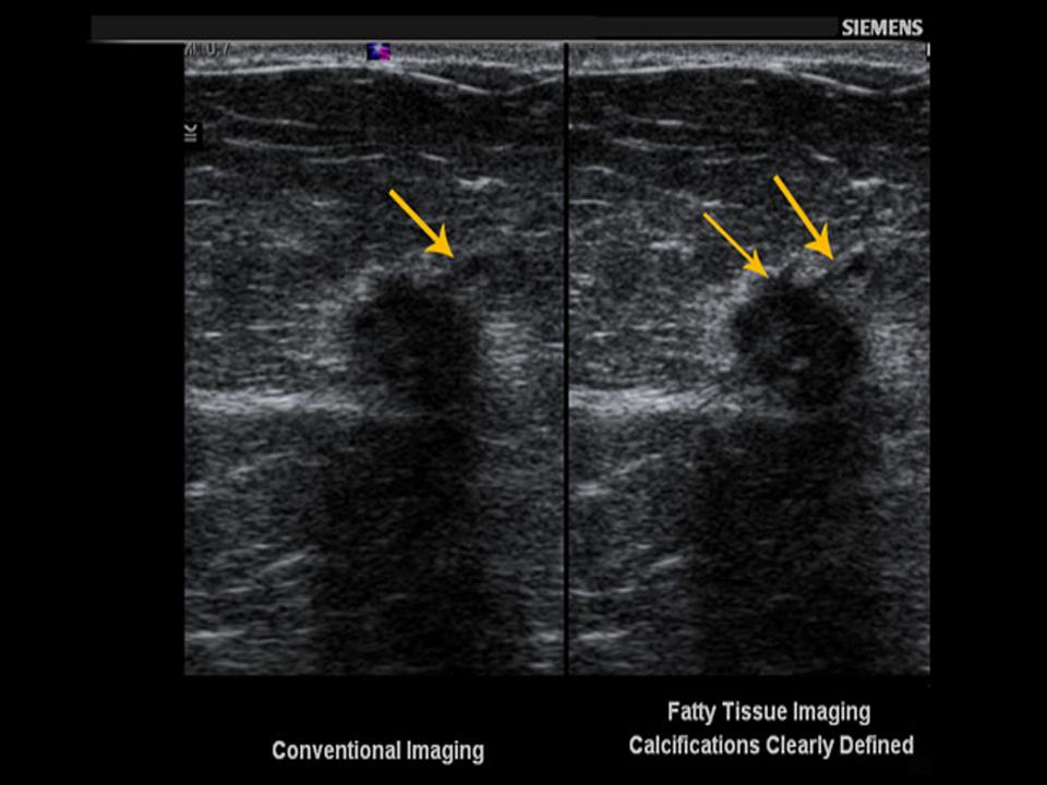
Diagnostics - Ultrasound

Improved detection sensitivity and characterization


Diagnostics - Mammography

Exact localization

Diagnostics - Magnetic Resonance

Excellent evaluation of malignant patterns


breast care diagnostics CT

Accurate tumor staging

Mammography

Diagnostics – Mammography

Exact localization – with Mammography

Diagnostics mammography
MammoTest’s polar coordinate approach offers precise positioning in all areas of the breast

In the diagnostic process, you need a solution for magnification and localization procedures, digital spot imaging, specimen radiography, galactography or image-guided stereotactic needle and core biopsies.

MAMMOMAT Inspiration
Stereotactic Biopsy with MAMMOMAT Inspiration

You may choose to perform diagnostic mammography and biopsies with one economical all-in-one solution. The MAMMOMAT Inspiration with stereotactic biopsy option features a fully integrated system and a highly automated workflow. The entire procedure can be controlled through the single compact-sized Acquisition Workstation that features the syngo® user interface. The biopsy unit of the Inspiration weight less than 11 pounds and is easy to handle. The needle holders are integrated into the biopsy tower. To prepare the system for biopsy, the unit is slid over the detector.

MammoTest
Eye contact with patient

Or you may decide on a dedicated biopsy solution placing the patient in the more relaxing prone position. With our biopsy system MammoTest™, procedures are performed out of patients’ view. At the same time, however, you can maintain eye contact with your patients, and have full patient and lesion access during the procedure, thanks to MammoTest’s 360° range of gantry movement. In addition, the “Target on Scout” functionality gives you the flexibility to use the 0° image for position calculation, thus potentially reducing the number of image retakes. And its lateral arm makes MammoTest ideally suited for small breasts.

Ultrasound

Diagnostics – Ultrasound

Improved detection sensitivity and characterization – with Ultrasound

eSie Touch elasticity
Qualitative information by demonstrating the typical internal characteristic pattern of these cysts

eSie Touch elasticity imaging
Our unique eSie Touch™ elasticity imaging is a new method that utilizes a real-time, dual imaging mode to demonstrate the simultaneous display of the standard B-mode image (composition of tissue) and the elastogram (relative stiffness) side-by-side. eSie Touch elasticity imaging is performed with a standard, high-frequency linear transducer and allows clinicians to see a new, different type of ultrasound data. The mechanical properties of tissue provide additional information enabling increased diagnostic findings and confidence.

Fatty tissue imaging

Fatty tissue imaging

Ultrasound techniques have historically used a fixed speed of sound for all imaging, despite the fact that the speed of sound varies in different types of tissue. Siemens Fatty Tissue Imaging technology uses an optimal speed-of-sound adaptation for fatty breast tissue which corrects for phase aberration, thus increasing detail and contrast resolution, and improving boundary detection. The results are better detection and identification of subtle lesion characteristics leading to increased diagnostic confidence. Fatty Tissue Imaging is a real-time, easy-to-use, single-button solution, which is applied during the standard ultrasound exam and requires no additional time to complete the study.

Magnetic Resonance

Diagnostics – Magnetic Resonance

Excellent evaluation of malignant patterns – with Magnetic Resonance

Dense breasts with MR

Dense breast tissue and breast implants are a major challenge when imaging breast cancer tumors in the diagnostic process; this is where Magnetic Resonance come into play. The diagnostic procedure includes not only the visualization and sizing but also the characterization and evaluation of whole-body lymph nodes. For staging and diagnosis, Magnetic Resonance offers ideal solutions. 3D imaging of the breast provides excellent information about lesions, normal and malignant tissues. All of this helps to determine the lesions extent and type.

MAGNETOM Espree Pink
MAGNETOM Espree - Pink

We have complemented our MAGNETOM family with a dedicated MR breast scanner: MAGNETOM Espree – Pink, the next generation of breast MRI. Whether for diagnostics, during therapy or follow-up, the new system offers unmatched patient comfort with Pink Comfort, unique applications with Pink Applications, and an innovative workflow with Pink Workflow. In short: MAGNETOM Espree – Pink combines the open-bore design of MAGNETOM Espree and a wide range of applications dedicated to improve diagnostic confidence with the Sentinelle Vanguard for Siemens solution that offers exceptional breast imaging and biopsy capabilities. The workplaces are equipped with a great number of computer-aided tools such as syngo® BreVis for effective breast reading including BIRADS reporting and syngo BreVis Biopsy for interventional procedure planning.

Whole body imaging with Tim
Whole-body image of patient with breast cancer and lung metastasis

Our MAGNETOM systems with the standard syngo® MR Breast Suite are ideal for imaging lesions in women with dense breasts and implant imaging: dedicated protocols and techniques can help visualize lesions and breast implants. In addition, they offer our unique Tim (Total imaging matrix) technology for pristine image quality. This means you'll have superior visualization. And, you'll obtain one of the highest signal-to-noise ratios with coverage from 5 mm up to 205 cm. With iPAT acceleration in all directions, Tim also delivers accelerated acquisition speed - while still offering the high spatial resolution needed for breast diagnosis.


Images courtesy of First Hill Diagnostic Imaging Center, Seattle, WA, USA

syngo GRACE

Metabolic information can be gained with syngo® GRACE, our application for quantitative MR breast spectroscopy. Having the ability to detect metabolic changes in tissue, it may provide additional confidence as a tool for the diagnosis of breast lesions. With choline acting as a biomarker, you can gain essential information on the biochemical composition of lesions.


This information can help to determine if lesions are malignant or not - to help reduce the patient risk of unnecessary breast biopsies as well as enable an accurate assessment of the efficacy of the treatment such as chemotherapy.
 

syngo VIEWS
Increased wash-in of an extensive ductal carcinoma in situ

Our software solution syngo® VIEWS provides both anatomical and functional information for exact tumor and lesion staging. By stimulating the water spins, it shows excellent fat suppression and provides 3D bilateral breast imaging in submilimeter range. You benefit from full coverage of both breasts and good lateral coverage up to the axilla region at both the highest temporal and highest spatial resolution. And combining syngo VIEWS with Parallel imaging's technique (syngo® GRAPPA), you also get superb protocols for high-resolution breast scanning. Due to its isotropic voxel size, images can be easily reformatted in any desired slice orientation.

CT

Diagnostics - Computed Tomography

Accurate staging - with Computed Tomography

Diagnostics CT

When staging a tumor, it is important to determine the presence of additional tumors and potential malignancies throughout the body. For this, it is important to have outstanding image quality with high spatial resolution and optimum coverage.

SOMATOM Definition AS

SOMATOM® Definition AS, the world’s first Adaptive Scanner, is a fundamentally new way to use single-source CT, provides a new way to use single source CT. Imagine a CT scanner that truly adapts to your patients and your clinical questions – making it an excellent imaging choice in virtually any clinical field. Opening up new worlds of clinical and economic possibilities. Increasing the quality and efficiency of imaging capabilities.

syngo CT Oncology
syngo CT Oncology

Our software syngo® CT Oncology provides unrivaled diagnostic confidence for routine tumor staging – especially for the evaluation of lesions in typical locations such as in the lung, liver or lymph nodes. Dedicated autopilots automate segmentation of user identified lesions, and measurement is manual, enabling robust calculation of the lesion size, including besides the standards RECIST (1.0) and WHO the forward-looking volume evaluation of the tumor, and the tumor burden. syngo CT Oncology uniquely offers a one-stop shop for high speed and confidence in staging.


“The Adaptive Scanner, SOMATOM Definition AS, enables us to address any clinical question in any situation.”

Joseph Schoepf, MD, Department of Radiology, Medical University of South Carolina, Charleston, USA
 

Subscription Button Icon
Be the first to know about our events, training, and news