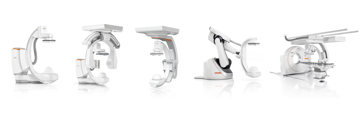
There’s a moment in every procedure when clarity changes everything. When uncertainty gives way to precision. When the right image unlocks the right decision.
The new ARTIS portfolio was built for that moment.
Imagine a unified portfolio designed to meet every clinical need, across every clinical segment, with uncompromising performance. Powered by the OPTIQ AI imaging chain and seamlessly integrated syngo software, this is more than a collection of systems. It’s a single, intelligent ecosystem engineered to deliver:
- consistent imaging
- intuitive workflows
- scalable innovation in every room.
Because when technology adapts to clinicians, possibilities expand. The ARTIS portfolio puts those possibilities within your reach.
OPTIQ AI
Clear insights. Powered by AI.
During minimally invasive procedures, it is critical to have a clear view of anatomies and devices. Yet complex imaging tasks or challenging patient conditions often impact image quality. OPTIQ AI delivers constant image quality1 defined by CNR in support of the ALARA principle, independent of patient or C-arm angulation. On top, an AI-powered algorithm reduces image noise in real time across different 2D imaging modes.
Make AI-powered imaging and clear insights your standard during interventions – with OPTIQ AI.
See for yourself: OPTIQ AI makes the difference
Neuro Interventions
04
Courtesy of Prof. René Chapot, MD, Alfried Krupp Krankenhaus Essen, Germany
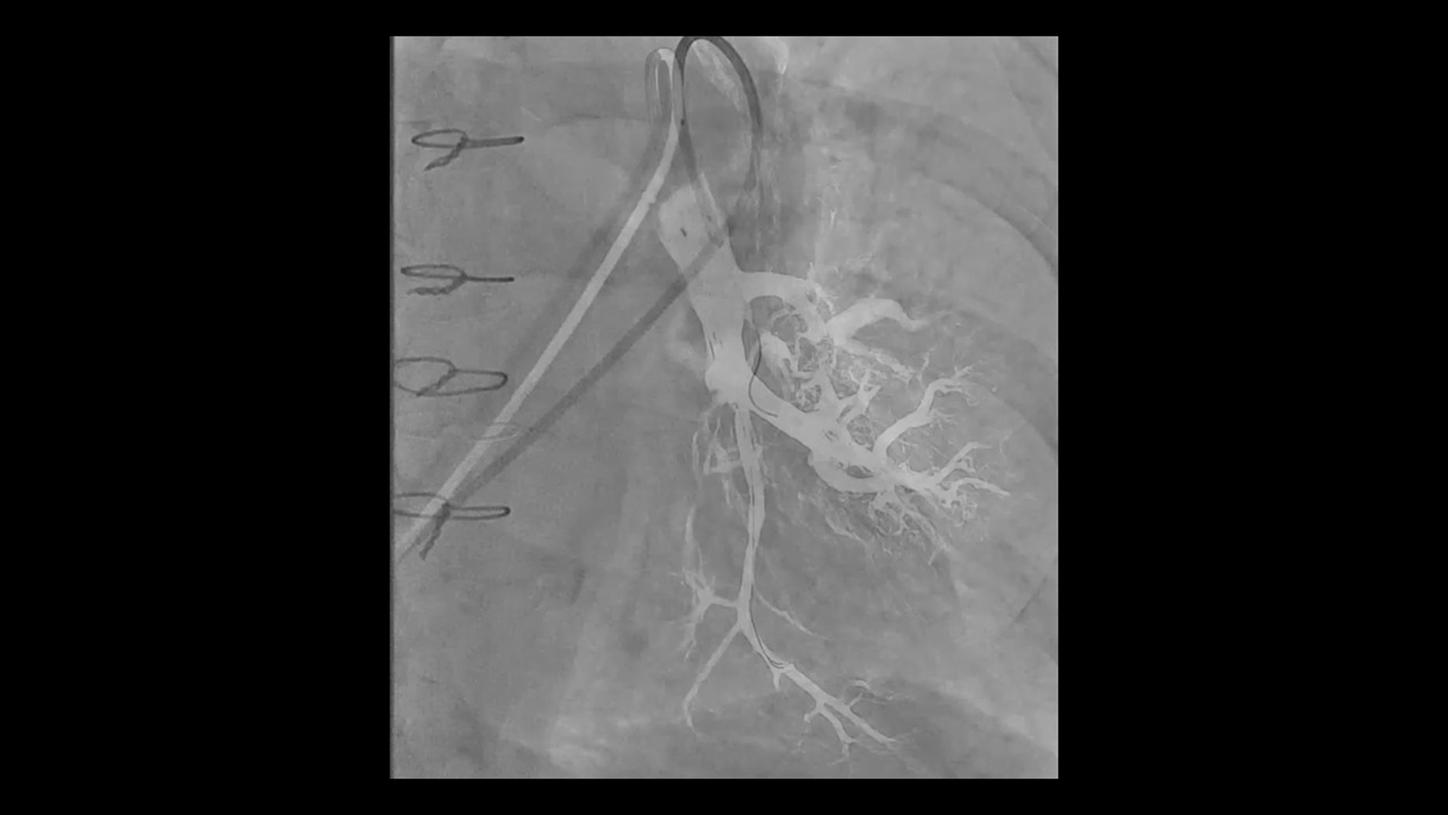
Visibility of small vessels and tissue perfusion.
Courtesy of Prof. Samuel Tobias Sossalla, MD, Kerckhoff-Klinik, Bad Nauheim, Germany
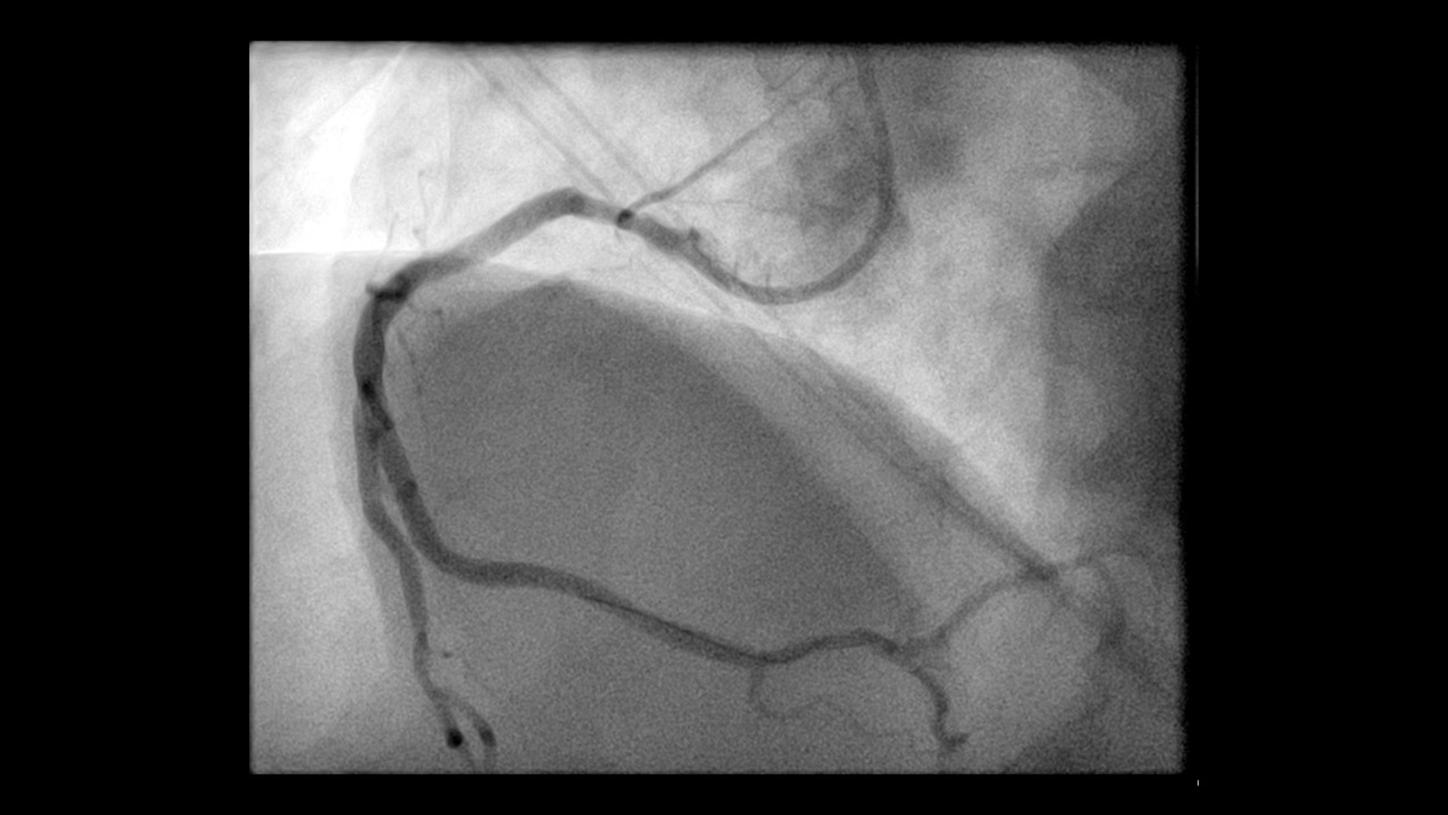
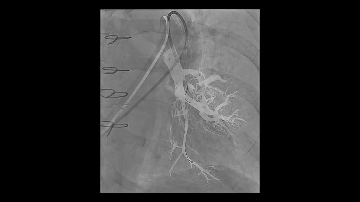
Percutaneous Coronary Intervention
AI-based denoising of acquisition scene of right coronary artery
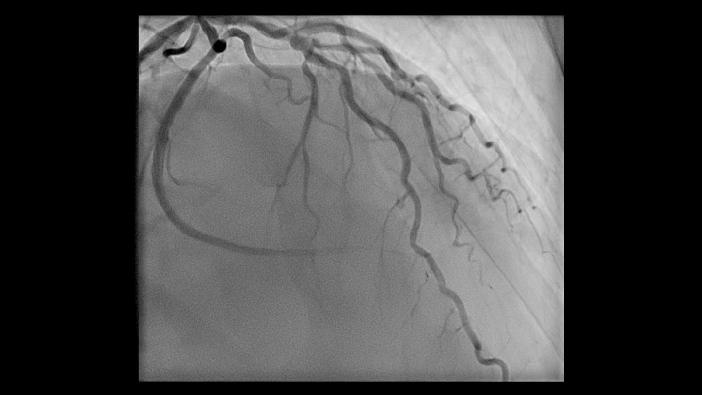
Courtesy of Prof. Samuel Tobias Sossalla, MD, Kerckhoff-Klinik, Bad Nauheim, Germany
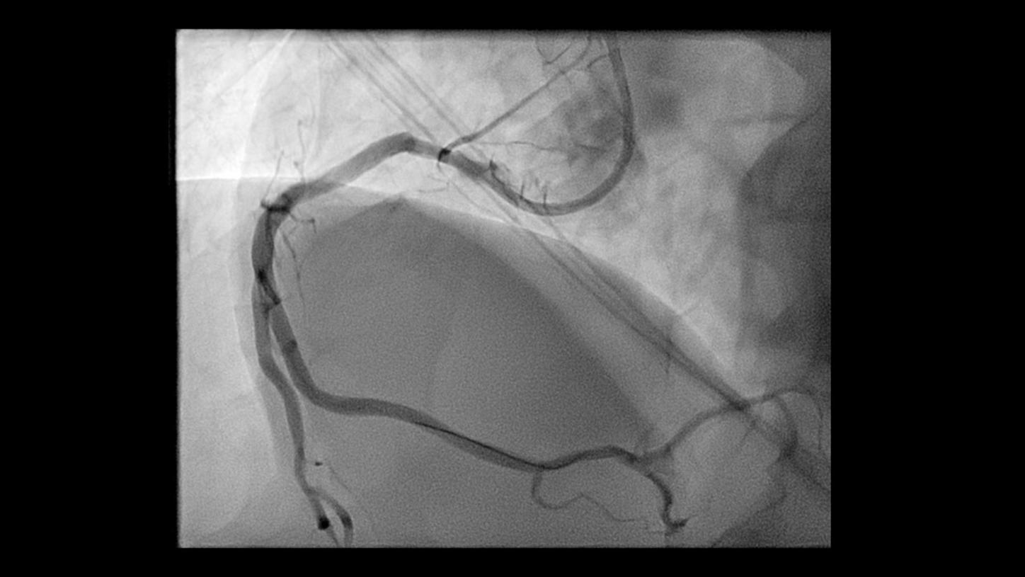
Percutaneous Coronary Intervention
AI-based denoising of acquisition images of left coronary artery
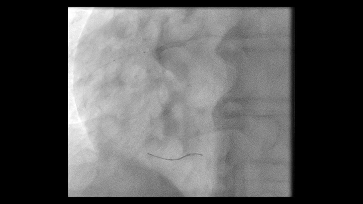
Courtesy of Prof. Samuel Tobias Sossalla, MD, Kerckhoff-Klinik, Bad Nauheim, Germany
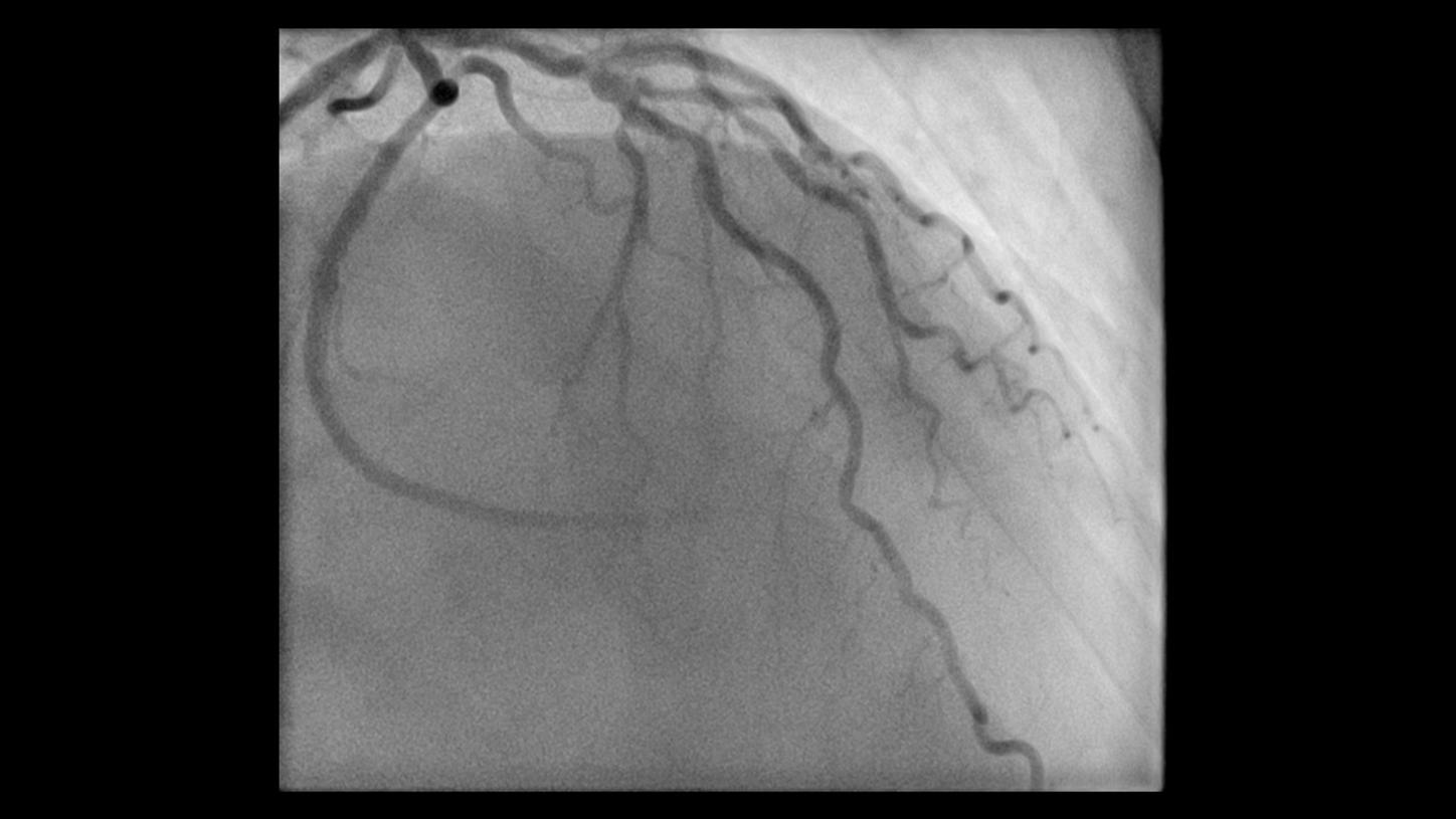
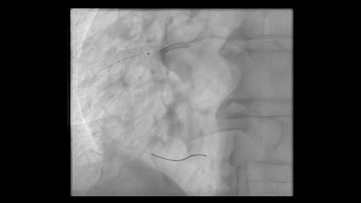
Percutaneous Coronary Intervention
AI-based denoising of fluoroscopy images
Courtesy of University Heart Center Freiburg/Bad Krozingen, Germany
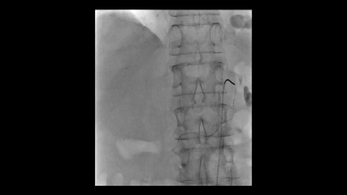
Constant image quality even in steep angulations.1
Even in steep angulations like spider view, OPTIQ AI delivers constant image quality defined by CNR in support of the ALARA principle.
Video 1
Courtesy of University Heart Center Freiburg/Bad Krozingen, Germany
Constant image quality even in steep angulations.1
Even in steep angulations like spider view, OPTIQ delivers constant image quality according to your pre-set image quality at lowest reasonable achievable dose.
Video 2
Courtesy of University Heart Center Freiburg/Bad Krozingen, Germany
Excellent device visibility at lowest reasonable achievable dose
Structure Scout enables optimization on specific materials, e.g., iron, tantalum, or platinum to save dose while maintaining excellent device visibility.4
Courtesy of Prof. Bernhard Meyer, MD, Hanover Medical School, Hanover, Germany
Selective internal radiotherapy
AI-based denoising of fluoroscopy scene
Courtesy of Prof. Bernhard Meyer, MD, Hanover Medical School, Hanover, Germany
Selective internal radiotherapy
AI-based image denoising together with overlay ref
Courtesy of Prof. Bernhard Meyer, MD, Hanover Medical School, Hanover, Germany
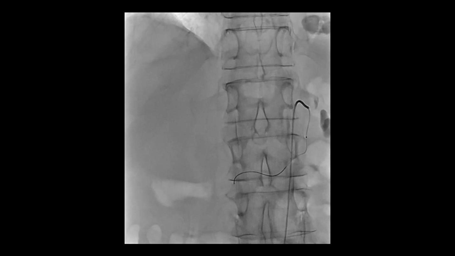
Transjugular intrahepatic portosystemic shunt
AI-based denoising of fluoroscopy images
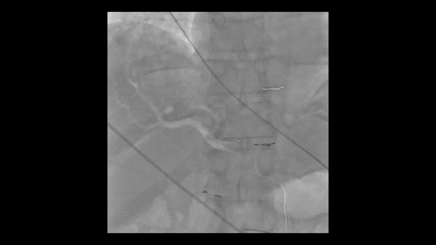
Courtesy of Prof. Bernhard Meyer, MD, Hanover Medical School, Hanover, Germany
Chemosaturation
AI-based image denoising together with overlay ref
Courtesy of Prof. Bernhard Meyer, MD, Hanover Medical School, Hanover, Germany
Pulmonary procedure
AI-based image denoising together with overlay ref

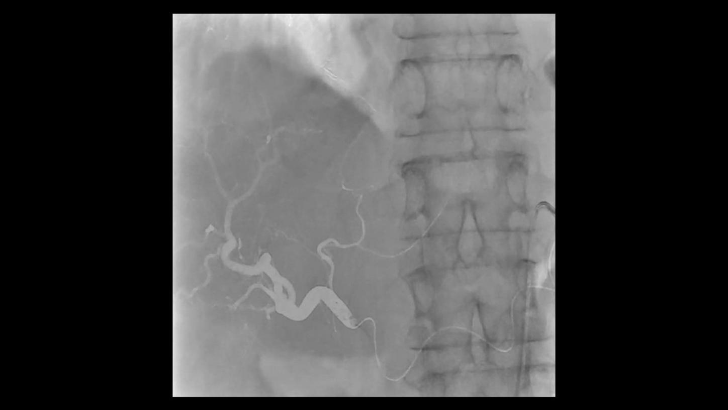
Courtesy of Prof. Florian Wolf, MD, Prof. Christian Loewe, MD, Allgemeines Krankenhaus Wien – Medical University Vienna, Austria
Prostate artery embolization (PAE)
Excellent visibility of tiny vessels with OPTIQ
Left side: DAP 15.38 µGy·m2/f
Right side: DAP 10.6 µGy·m2/f
 Without Structure Scout
 With Structure Scout
Courtesy of Prof. Bernhard Meyer, MD, Hanover Medical School, Germany
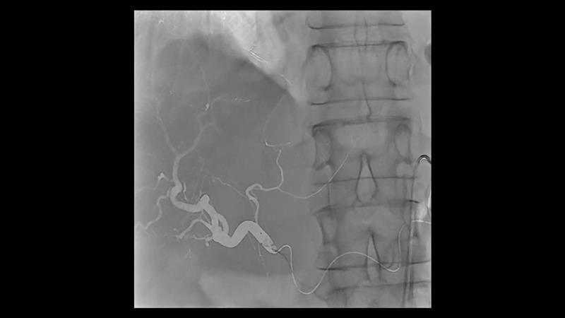
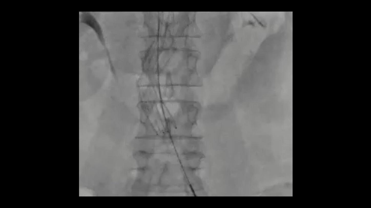
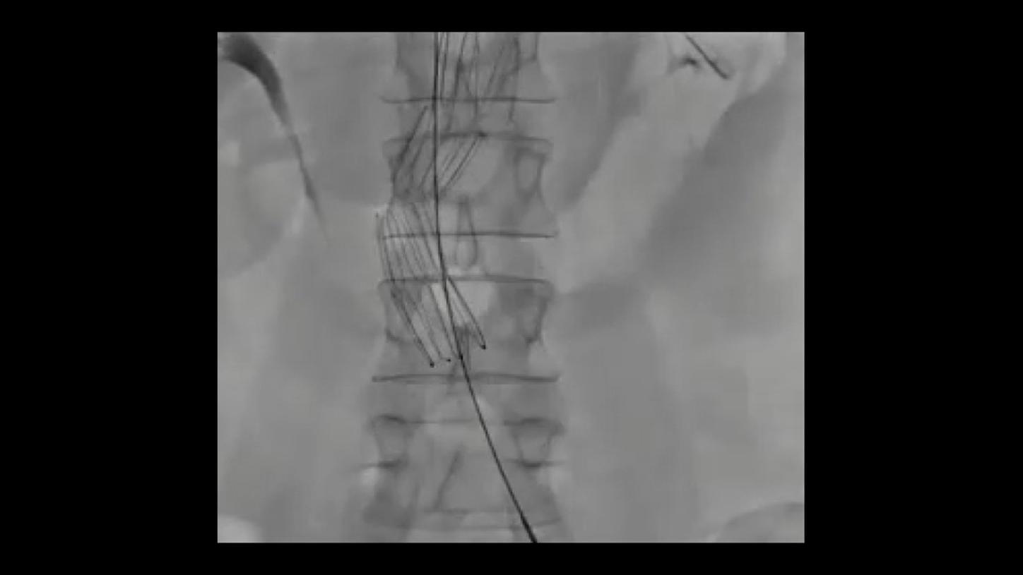
TIPS procedure using a contrast-filled balloon over an iron wire
47-year-old female, BMI 22.5
Less dose at same device visibility with Structure Scout
Left side: DAP: 0.63 µGy·m2/f, without Structure Scout
Right side: DAP: 0.45 µGy·m2/f, with Structure Scout

Courtesy of Prof. Dierk Vorwerk, MD, Institute of Radiology, Klinikum Ingolstadt, Germany
Visualization of fine structures and compensation for motion artifacts
“What is very impressive is the detailed clarity of the images and the image resolution that is gorgeous.” 5
Prof. Dierk Vorwerk, MD
Head of the Department of Radiology,
General Hospital, Ingolstadt, Germany

Courtesy of Prof. Florian Wolf, MD, Prof. Christian Loewe, MD, Allgemeines Krankenhaus Wien – Medical University Vienna, Austria
Excellent visibility of tiny vessels in the knee
Visualizing embolic material in fluoroscopy.
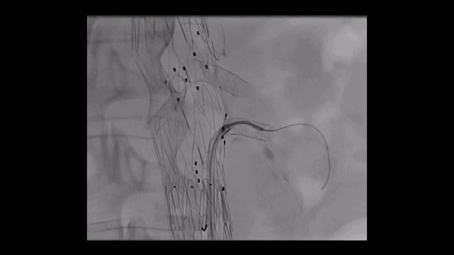
Courtesy of Prof. Markus Steinbauer, MD, Krankenhaus Barmherzige Brüder, Regensburg, Germany
Endoleak Repair
AI-based denoising of fluoroscopy images

Courtesy of Prof. Markus Steinbauer, MD, Krankenhaus Barmherzige Brüder, Regensburg, Germany
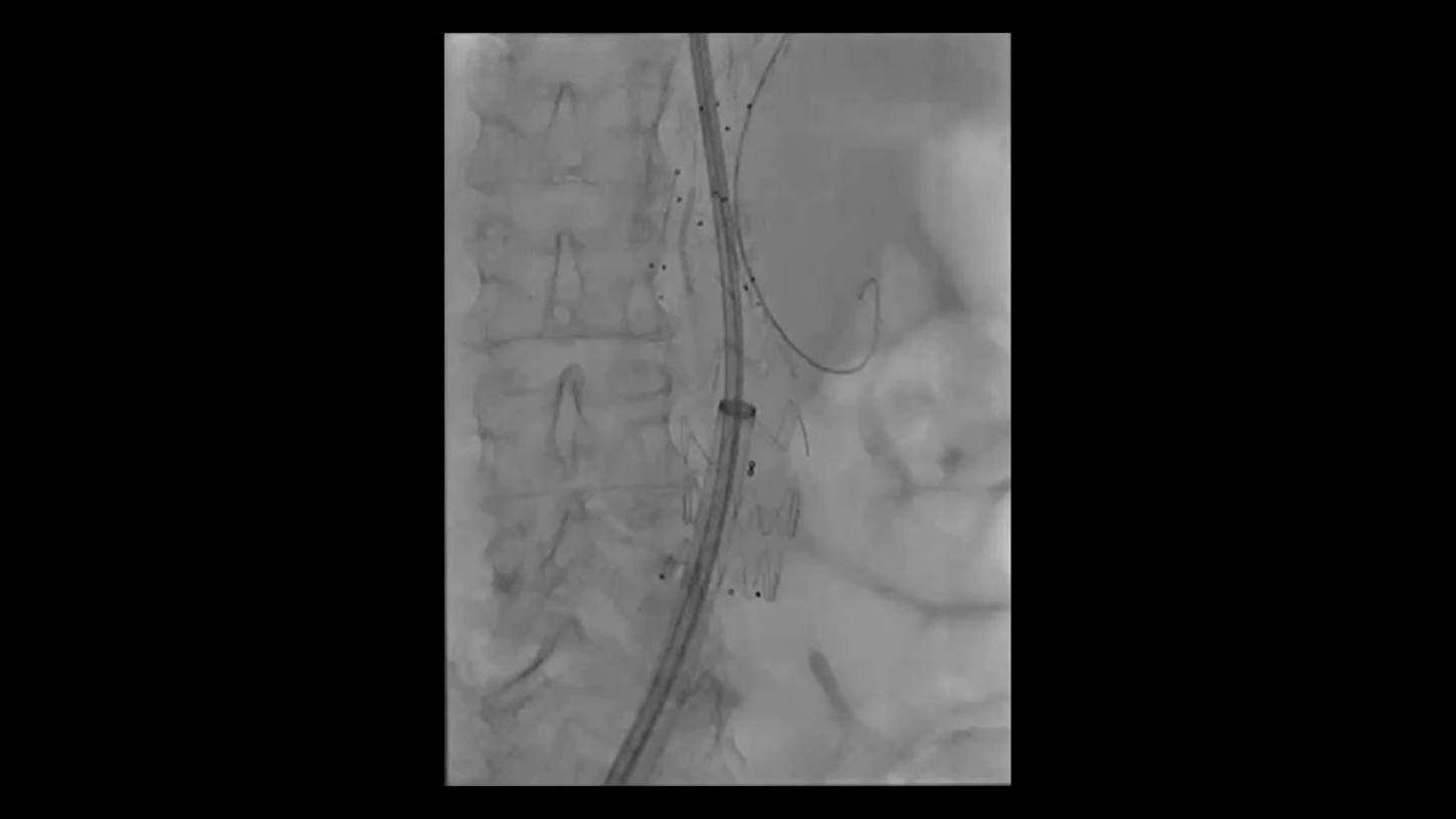
Fenestrated Endovascular Aortic Repair
AI-based denoising of fluoroscopy images
Courtesy of Prof. Markus Steinbauer, MD, Krankenhaus Barmherzige Brüder, Regensburg, Germany
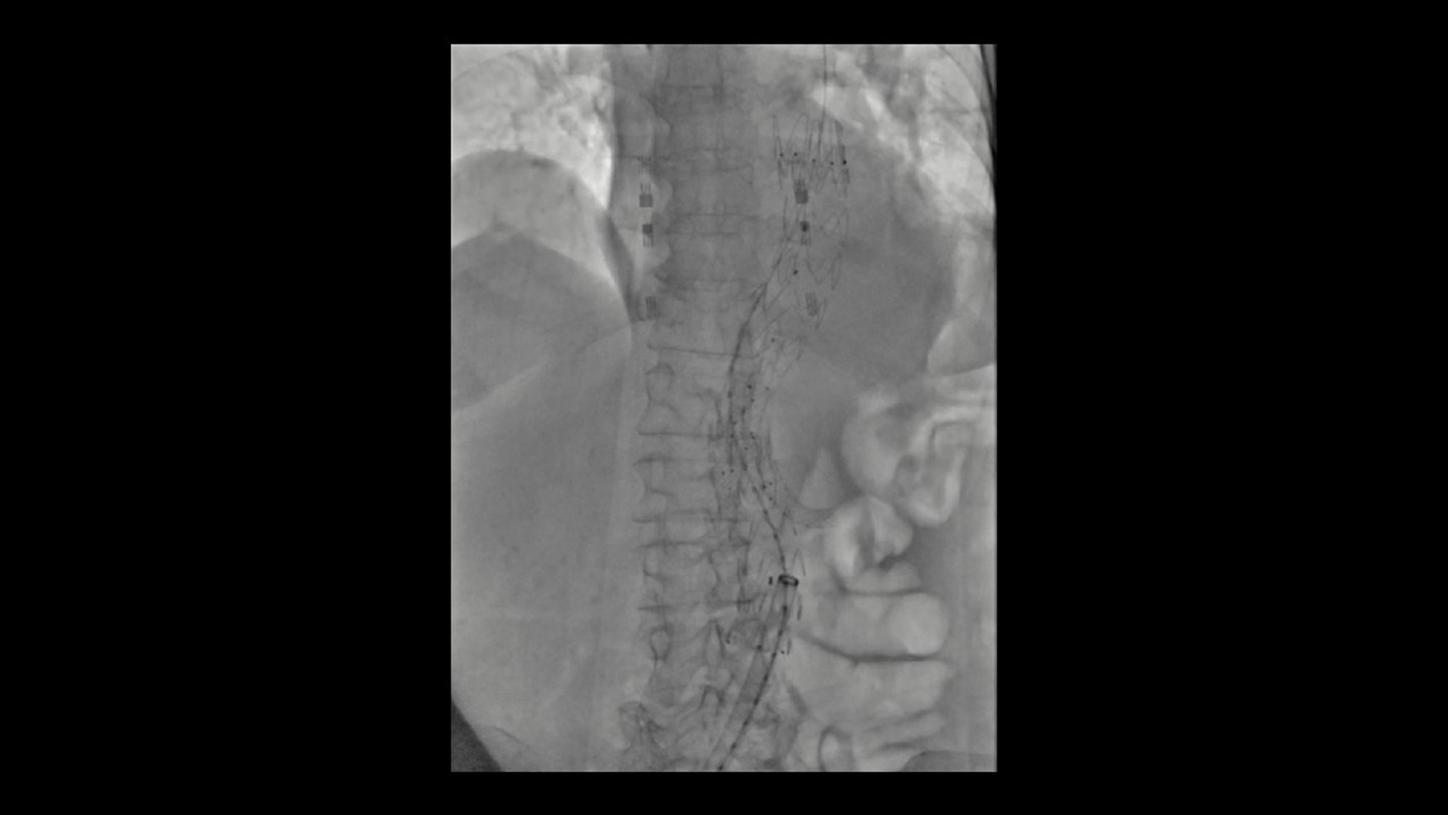
Fenestrated Endovascular Aortic Repair
AI-based denoising of fluoroscopy images
Courtesy of Prof. Markus Steinbauer, MD, Krankenhaus Barmherzige Brüder, Regensburg, Germany
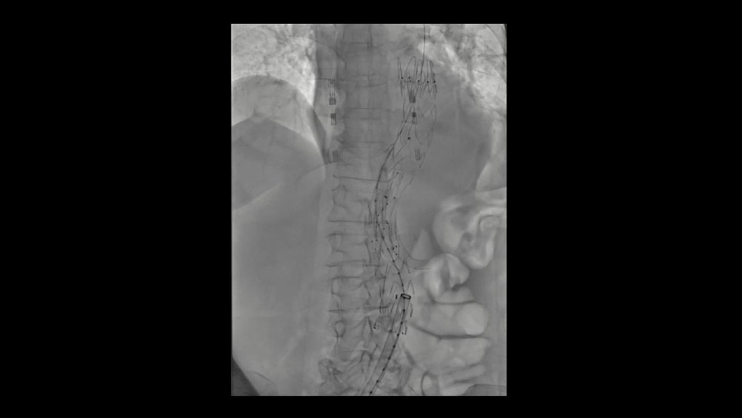
Thoracic Endovascular Aortic Repair
AI-based denoising of fluoroscopy scene

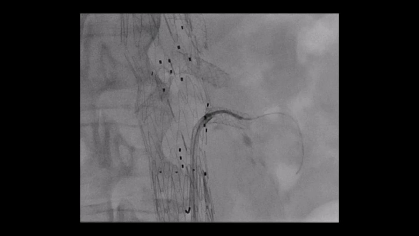
Courtesy of Prof. René Chapot, MD, Alfried Krupp Krankenhaus Essen, Germany
OPTIQ Roadmap: Significant improvements of device contrast over vessel map
“Impressive is the quality of the Fluoroscopy and the quality of the Roadmap. Increased image quality goes together with a reduction in doses.”5
Prof. René Chapot, MD
Head of the Department of Neuroradiology,
Alfried Krupp Hospital, Essen, Germany
 Without Structure Scout
 With Structure Scout
Courtesy of Prof. Bernhard Meyer, MD, Hanover Medical School, Hanover, Germany
AVM treatment using embolic material containing tantalum
32-year-old male patient, BMI 24
Less dose at same device visibility with Structure Scout
Left side: DAP: 0.15 µGy·m2/f, without Structure Scout
Right side: DAP: 0.026 µGy·m2/f, with Structure Scout

Courtesy of Jan Gralla, MD, Department of Neuroradiology, Inselspital, Bern, Switzerland
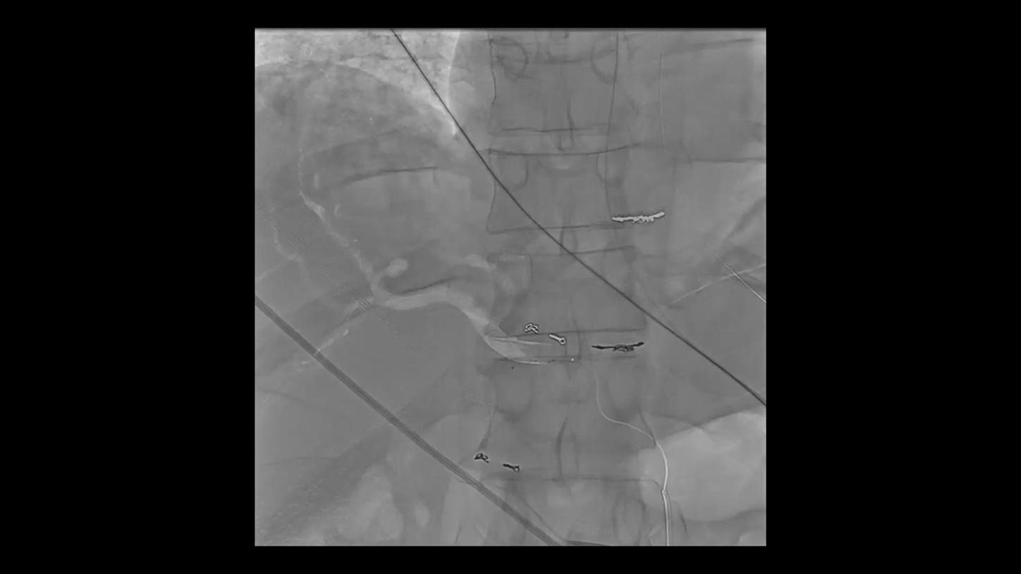
Visibility of stent struts
Neuro device protocol used for treatment success documentation after carotid artery stenting
Left side: Ballons of Mo.Ma Device
Right side: "Protegé"-Stent (10/7 x 30 mm)
Courtesy of Prof. René Chapot, MD, Alfried Krupp Krankenhaus Essen, Germany
Visibility of small vessels and tissue perfusion.
Courtesy of Prof. Samuel Tobias Sossalla, MD, Kerckhoff-Klinik, Bad Nauheim, Germany
Percutaneous Coronary Intervention
AI-based denoising of acquisition scene of right coronary artery
Courtesy of Prof. Samuel Tobias Sossalla, MD, Kerckhoff-Klinik, Bad Nauheim, Germany
Percutaneous Coronary Intervention
AI-based denoising of acquisition images of left coronary artery
Courtesy of Prof. Samuel Tobias Sossalla, MD, Kerckhoff-Klinik, Bad Nauheim, Germany
Percutaneous Coronary Intervention
AI-based denoising of fluoroscopy images
Courtesy of University Heart Center Freiburg/Bad Krozingen, Germany
Constant image quality even in steep angulations.1
Even in steep angulations like spider view, OPTIQ AI delivers constant image quality defined by CNR in support of the ALARA principle.
Video 1
Courtesy of University Heart Center Freiburg/Bad Krozingen, Germany
Constant image quality even in steep angulations.1
Even in steep angulations like spider view, OPTIQ delivers constant image quality according to your pre-set image quality at lowest reasonable achievable dose.
Video 2
Courtesy of University Heart Center Freiburg/Bad Krozingen, Germany
Excellent device visibility at lowest reasonable achievable dose
Structure Scout enables optimization on specific materials, e.g., iron, tantalum, or platinum to save dose while maintaining excellent device visibility.4
Courtesy of Prof. Bernhard Meyer, MD, Hanover Medical School, Hanover, Germany
Selective internal radiotherapy
AI-based denoising of fluoroscopy scene
Courtesy of Prof. Bernhard Meyer, MD, Hanover Medical School, Hanover, Germany
Selective internal radiotherapy
AI-based image denoising together with overlay ref
Courtesy of Prof. Bernhard Meyer, MD, Hanover Medical School, Hanover, Germany
Transjugular intrahepatic portosystemic shunt
AI-based denoising of fluoroscopy images
Courtesy of Prof. Bernhard Meyer, MD, Hanover Medical School, Hanover, Germany
Chemosaturation
AI-based image denoising together with overlay ref
Courtesy of Prof. Bernhard Meyer, MD, Hanover Medical School, Hanover, Germany
Pulmonary procedure
AI-based image denoising together with overlay ref
Courtesy of Prof. Florian Wolf, MD, Prof. Christian Loewe, MD, Allgemeines Krankenhaus Wien – Medical University Vienna, Austria
Prostate artery embolization (PAE)
Excellent visibility of tiny vessels with OPTIQ
Left side: DAP 15.38 µGy·m2/f
Right side: DAP 10.6 µGy·m2/f
Without Structure ScoutWith Structure ScoutCourtesy of Prof. Bernhard Meyer, MD, Hanover Medical School, Germany
TIPS procedure using a contrast-filled balloon over an iron wire
47-year-old female, BMI 22.5
Less dose at same device visibility with Structure Scout
Left side: DAP: 0.63 µGy·m2/f, without Structure Scout
Right side: DAP: 0.45 µGy·m2/f, with Structure Scout
Courtesy of Prof. Dierk Vorwerk, MD, Institute of Radiology, Klinikum Ingolstadt, Germany
Visualization of fine structures and compensation for motion artifacts
“What is very impressive is the detailed clarity of the images and the image resolution that is gorgeous.” 5
Prof. Dierk Vorwerk, MD
Head of the Department of Radiology,
General Hospital, Ingolstadt, Germany
Courtesy of Prof. Florian Wolf, MD, Prof. Christian Loewe, MD, Allgemeines Krankenhaus Wien – Medical University Vienna, Austria
Excellent visibility of tiny vessels in the knee
Visualizing embolic material in fluoroscopy.
Courtesy of Prof. Markus Steinbauer, MD, Krankenhaus Barmherzige Brüder, Regensburg, Germany
Endoleak Repair
AI-based denoising of fluoroscopy images
Courtesy of Prof. Markus Steinbauer, MD, Krankenhaus Barmherzige Brüder, Regensburg, Germany
Fenestrated Endovascular Aortic Repair
AI-based denoising of fluoroscopy images
Courtesy of Prof. Markus Steinbauer, MD, Krankenhaus Barmherzige Brüder, Regensburg, Germany
Fenestrated Endovascular Aortic Repair
AI-based denoising of fluoroscopy images
Courtesy of Prof. Markus Steinbauer, MD, Krankenhaus Barmherzige Brüder, Regensburg, Germany
Thoracic Endovascular Aortic Repair
AI-based denoising of fluoroscopy scene
Courtesy of Prof. René Chapot, MD, Alfried Krupp Krankenhaus Essen, Germany
OPTIQ Roadmap: Significant improvements of device contrast over vessel map
“Impressive is the quality of the Fluoroscopy and the quality of the Roadmap. Increased image quality goes together with a reduction in doses.”5
Prof. René Chapot, MD
Head of the Department of Neuroradiology,
Alfried Krupp Hospital, Essen, Germany
Without Structure ScoutWith Structure ScoutCourtesy of Prof. Bernhard Meyer, MD, Hanover Medical School, Hanover, Germany
AVM treatment using embolic material containing tantalum
32-year-old male patient, BMI 24
Less dose at same device visibility with Structure Scout
Left side: DAP: 0.15 µGy·m2/f, without Structure Scout
Right side: DAP: 0.026 µGy·m2/f, with Structure Scout
Courtesy of Jan Gralla, MD, Department of Neuroradiology, Inselspital, Bern, Switzerland
Visibility of stent struts
Neuro device protocol used for treatment success documentation after carotid artery stenting
Left side: Ballons of Mo.Ma Device
Right side: "Protegé"-Stent (10/7 x 30 mm)
Courtesy of Prof. René Chapot, MD, Alfried Krupp Krankenhaus Essen, Germany
Visibility of small vessels and tissue perfusion.
Without Structure ScoutWith Structure ScoutWithout Structure ScoutWith Structure ScoutThe new ARTIS portfolio