World-class Speakers from Around the Globe
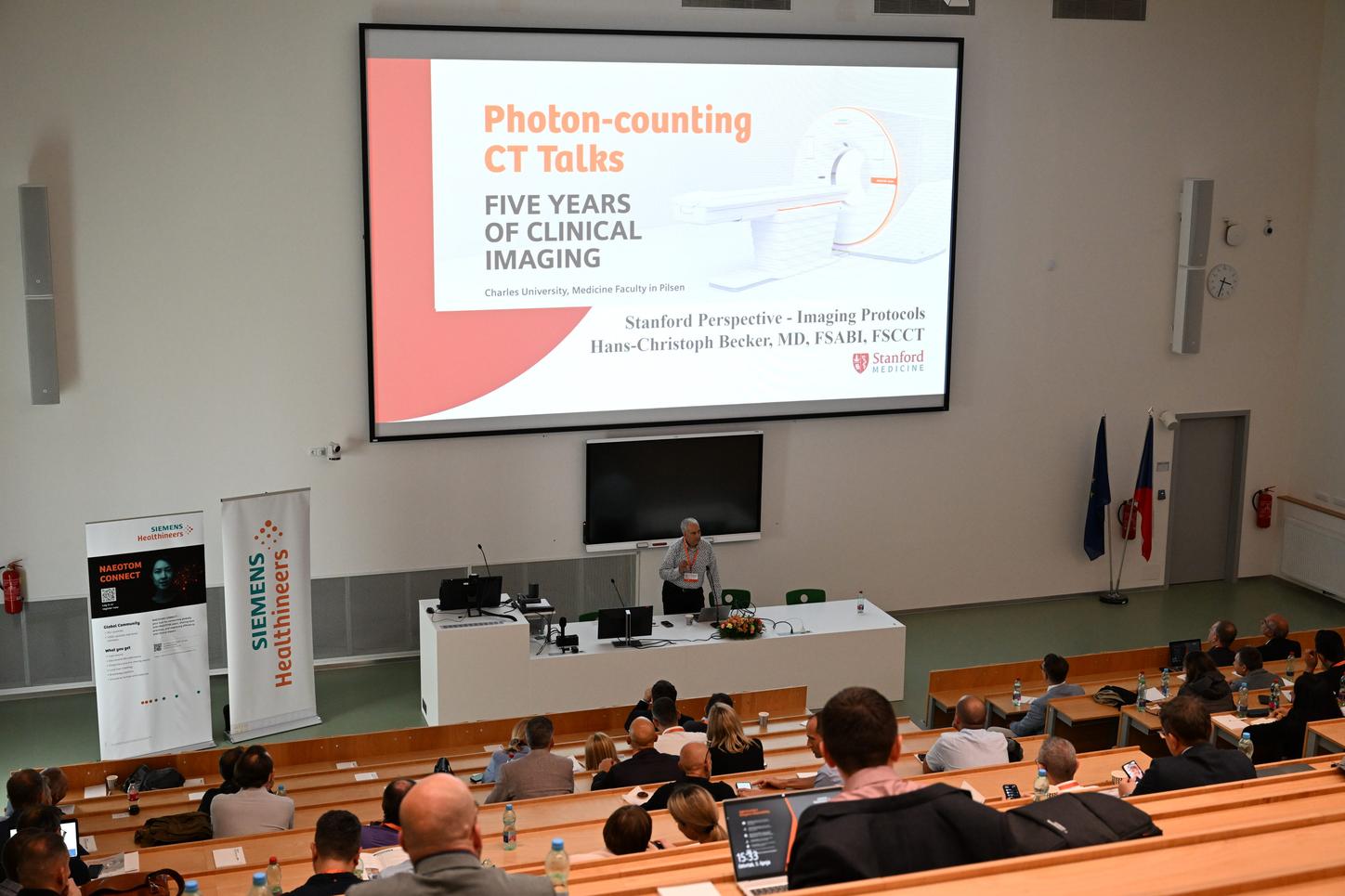
The two-day gathering, held on 5–6 June 2025, provided a comprehensive look at the evolution and practical impact of PCCT in clinical settings. Leading radiologists from institutions such as Stanford University, the University of Tübingen, Maastricht UMC+, and University Hospital Zurich shared their experiences, along with key figures from Czech radiology, notably from the University Hospital in Pilsen. The programme covered the use of PCCT in cardiology, oncology, neurology, paediatrics, as well as the integration of artificial intelligence into diagnostic processes.
A Technological Breakthrough and the Role of Siemens Healthineers
The conference was held under the patronage of Siemens Healthineers, the manufacturer of the NAEOTOM Alpha scanners – the world’s first photon-counting CT systems approved for clinical use. Thomas Flohr of Siemens Healthineers recalled that as early as 2001, it was clear that PCCT had the potential to bring a qualitative shift in CT imaging. The development process spanned nearly twenty years, from fundamental research to clinical prototypes and finally to routine clinical deployment.
Bernhard Schmidt, Head of CT Research and Development at Siemens Healthineers, likened the difference between conventional CT and PCCT to comparing a blurry photo booth snapshot to a sharp image taken with a professional DSLR camera. The technology delivers images with higher resolution, lower noise, and significantly improved detail readability, while also providing spectral data with every scan.
Insights from Pilsen and Beyond
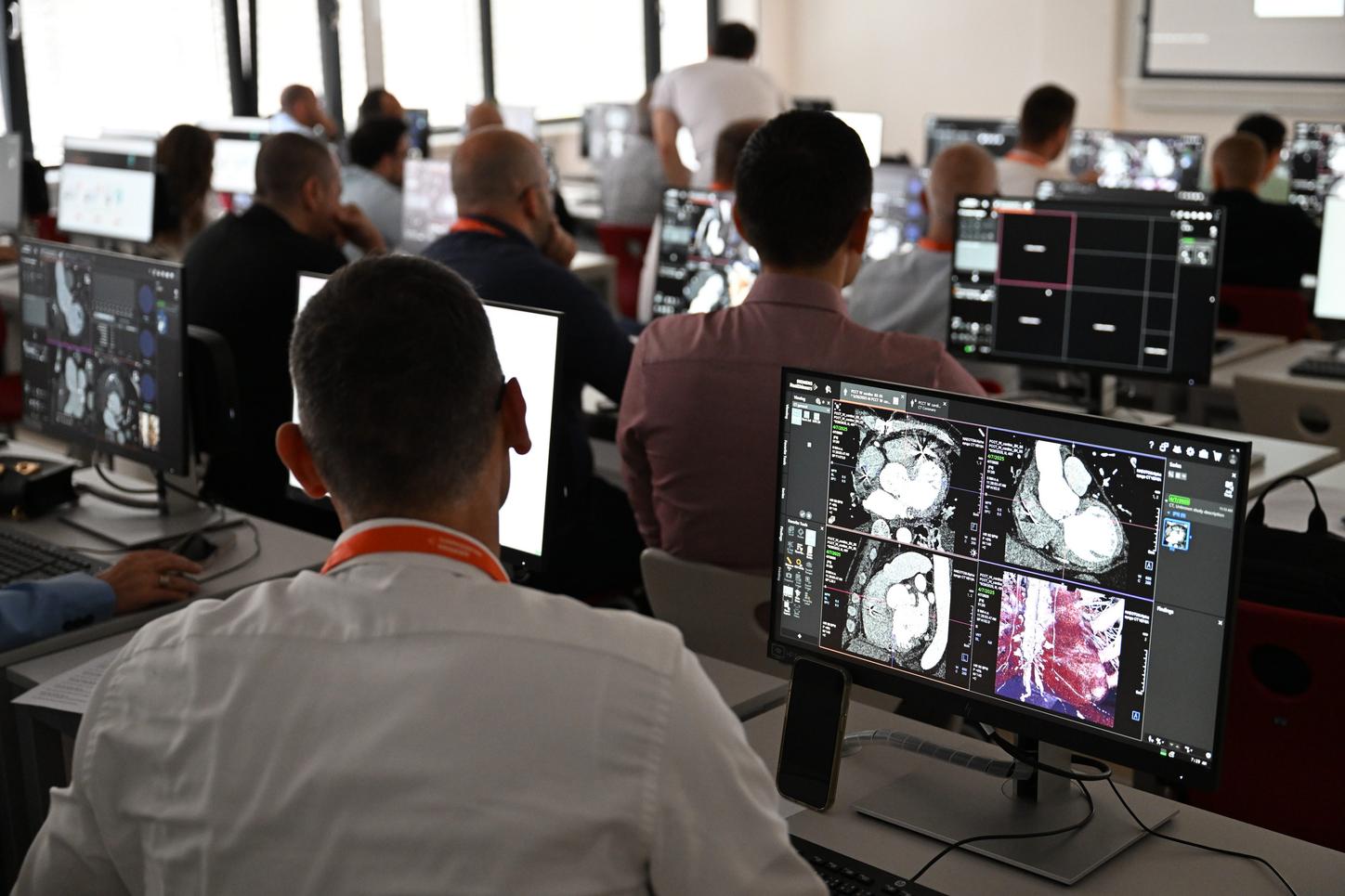
Among those presenting practical insights was Professor Jiří Ferda, MD, PhD, from the University Hospital in Pilsen, along with his team. The hospital currently operates three models from the NAEOTOM Alpha series – Alpha.Peak, Alpha.Pro and Alpha.Prime – which are used across emergency departments, paediatric radiology, intensive care, as well as urology and pulmonology units.
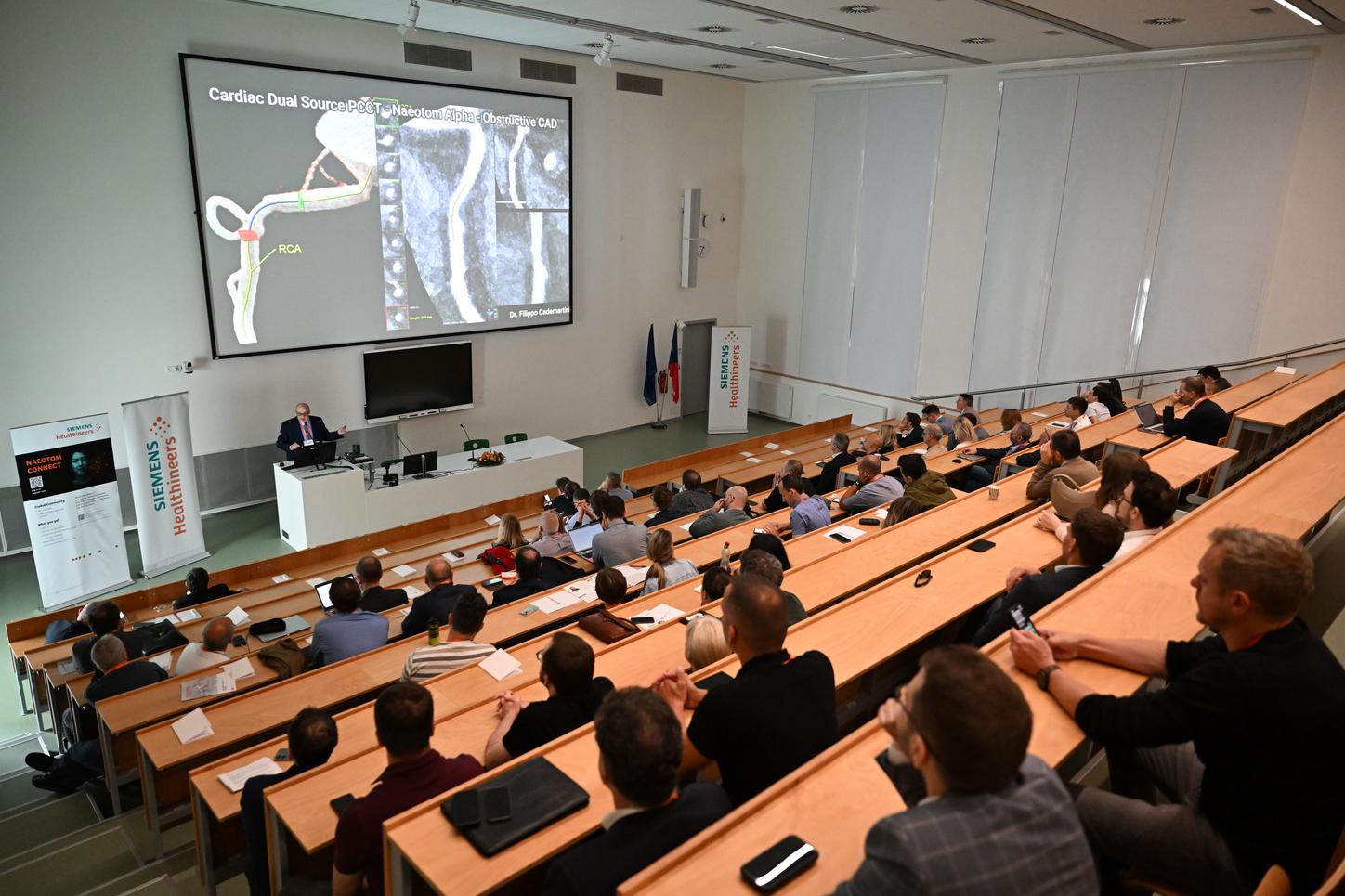
A significant portion of the programme focused on the clinical applications of PCCT – for example in imaging of the brain, pelvis, pancreas, spine, and prostate. According to Professor Christoph Becker from Stanford, the technology has proven particularly effective in body imaging. Andreas Brendlin from Tübingen highlighted its benefits in oncology and pointed to another major advantage: a markedly reduced need for contrast agents. Lower doses mean reduced strain on the kidneys and other organs, while image quality remains crisp and clear – a crucial benefit for at-risk and oncology patients.
Photon-counting CT is Transforming Radiology
From the opening lectures to expert panel discussions, it became clear that photon-counting CT has not only entered clinical practice successfully but is also reshaping the approach to diagnosis. Thanks to PCCT, radiology is becoming more accurate, patient-friendly and predictive – and as echoed by many speakers, it is highly likely that this technology will become the new standard in CT imaging in the coming years.