
For clinicians and researchers in the department of
nuclear medicine at Switzerland’s Inselspital, Bern University Hospital, being
the first site to install Biograph Vision Quadra™ marked the conclusion of a
process that began when their team crossed the Atlantic to consult with Siemens
Healthineers as they designed the next-generation whole-body PET/CT scanner.
Photography by Benedikt Schnermann
Data courtesy of Inselspital, Bern, Switzerland
Listen to a dynamic ten-minute reading of the article.
Throughout this process, they shared their ideas and aspirations for how the scanner, which combines an extended axial field of view (FoV) with industry-leading detector technology, could advance routine clinical care and help answer new research questions. The installation of the scanner in October 2020 ushered in a new era for a department that is already recognized as a leader in the innovative use of PET/CT.
“This
is, in our opinion, a kind of a quantum leap of whole-body PET imaging,” says
Axel Rominger, MD, PhD, the department’s director and chief nuclear medicine
physician. “We are convinced this new technology will broaden our clinical horizons
and also, since we are a university hospital, our scientific frontiers with
manifold new opportunities.”
Maximum flexibility for clinical care
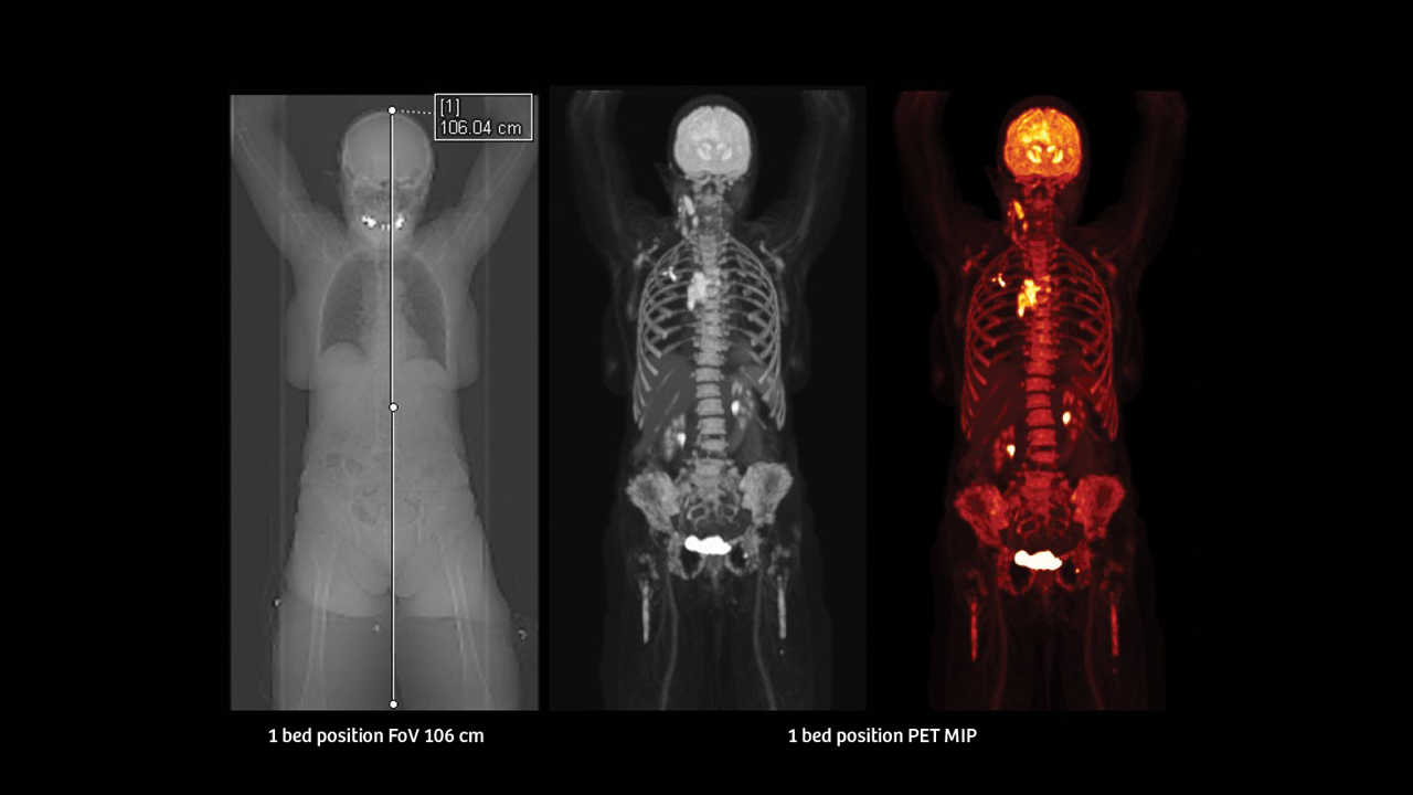
Rominger says that several features made the scanner an attractive choice. With four silicone photomultiplier (SiPM) detector blocks placed in series, the system offers a 106 cm axial FoV that enables whole-body imaging and results in a high effective sensitivity that helps make small lesions readily identifiable.
The
range of coverage enables physicians to image the brain, heart, and the entire
spinal cord in one pass, as opposed to several small passes that are later
pieced together via post-processing software. As an example, Rominger notes how
this could enable the diagnosis of neurodegenerative disorders, as well as
cardiac amyloidosis, in one scan and facilitates the identification of distant
metastases.
He explains how the enhanced sensitivity can allow PET/CT scans acquired with tracers of low positron activity, such as Yttrium-90 (90Y, commonly referenced as Y90 scans), to potentially be used for diagnostic purposes in addition to their already commonplace therapeutic use.
Rominger emphasizes that
Biograph Vision Quadra gives physicians a greater ability to limit radiation
dose or scan times depending on the specific needs of the patient. Minimizing
dose is particularly important for pediatric patients, while patients in pain
or with claustrophobia benefit from rapid scan times. “It’s flexible in two
directions, radiation burden and acquisition time,” he says. “This is
beneficial for the patient and also beneficial for our resources.”
“We are convinced this new technology will broaden our clinical horizons and also, since we are a university hospital, our scientific frontiers with manifold new opportunities.”

Bern University Hospital
Head radiology technician HF, Marco Viscione, notes that the system allows for the reduction of scan times to between four and six minutes for a typical head-to-hip scan. If circumstances warrant, scan times can be reduced below two minutes.
With a reduced scan time, patients can potentially spend less time in the clinic, and the clinic itself can perform more scans in a given day. At Inselspital, they can now easily perform more than 100 scans per week on their newest PET/CT.
Viscione adds that the increased patient throughput has allowed Inselspital to allocate a specific day each week to use the scanner for research purposes. Previously, research had to be conducted in the evenings after clinical scans were complete. Now, several research projects that seek to push image quality to new levels, while reducing dose and expanding the scope and utility of PET/CT, are underway.
“This super scanner needs a super team, so a single institute is not sufficient.”

Bern University Hospital
A new era for research
Kuangyu Shi, PhD, head of the Artificial Intelligence and Translational Theranostics Lab in the department of nuclear medicine, says the system’s expanded axial FoV and high sensitivity create new research opportunities in precision pharmacology and precision medicine. “With those things combined together, it’s not a small step further—I think it’s a breakthrough,” he says. “It opens up a new era.”
He and Rominger note the expanded axial FoV can give researchers greater insight into the pharmacokinetics of all major organs. Shi and his colleagues are working to develop parametric imaging algorithms to provide more meaningful quantitative data. “We now have precise and more systematic pharmacokinetics,” Shi says. “It enables systematic investigation of the interrelations of multiple organs.”

The patient in the clinical images within this article was scanned with a Biograph Vision Quadra, 5.1 mCi (191 mBq), 90-minute post-injection delay. CT: 100kV, 6 ref mAs.
In addition to aiding the development of new drugs and potentially providing prognostic information based on the uptake of a radiopharmaceutical in a patient, the ability to acquire whole-body dynamic PET images allows clinicians to follow tracer kinetics. The ability to acquire dynamic images over the whole body in one bed position is an essential component of a study in which Rominger is investigating the uptake of a radiotracer in the brain and spinal cord over time. “Dynamic imaging has great potential to really tell us what’s going on with the tracer within the body,” he says.
The sheer quantity of data generated by dynamic scans presents its own challenges, and Shi is developing reconstruction algorithms that seek to ease the identification of clinically relevant information. He is also working on reconstruction algorithms that have the potential to improve image quality even further. Beyond these algorithms, Shi and his colleagues are exploring opportunities for CT dose-reduction techniques.

He notes that artificial intelligence is not the only tool that he and his colleagues are using, but he emphasizes that it plays a critical role. With further dose reductions, he adds, opportunities for the use of PET in screening become feasible, and the monitoring of responses to treatments can be expanded.
“By hardware we have
already achieved a reduction of dose, maybe 10 times,” Shi says. “So if we can
further reduce the dose with algorithms, maybe another five times, this will be
fantastic.”
Expanding collaborations
Inselspital is building collaborations with pharmaceutical partners, and its team already includes experts in computer science and artificial intelligence. Shi says that broader collaborations are in order.
“We are now establishing more collaborations with our colleagues and even some outside the nuclear medicine community,” he says. “We believe that’s the only way to fully explore the potential of this super scanner. This super scanner needs a super team, so a single institute is not sufficient.”
“We had a lot of help from Siemens Healthineers and from the factory. Every week we had a meeting so we could see what software updates will come and what’s important for us.”

Bern University Hospital
Viscione adds that the sense of collaboration he and his colleagues experienced with the U.S.-based research and development team at Siemens Healthineers in Knoxville, Tennessee, continued after the scanner was installed, and it continues to this day.
“We had a lot of help from Siemens Healthineers and from the factory,” he says. “Every week we had a meeting so we could see what software updates will come and what’s important for us. For me, it’s important to speak with engineering, and I think this is really helpful for both sides.”
The scanner has proven to
be a workhorse in clinical routine while it also supports Inselspital’s mission
of advancing the future of clinical care through research. “This is our job and
duty: to push the limits further and further,” Rominger says. “And so, in the
future, things can become even better.”
About the author
Sameh Fahmy, MS, is an award-winning freelance medical and technology journalist based in Athens, Georgia, USA.