Get further. With True Dual Energy.DEfinitely Siemens Healthineers

Dual Energy Heart PBV

More and more radiologists are relying on the rich diagnostic possibilities offered by True Dual Energy imaging on Siemens Healthineers' CT scanner fleet ranging from SOMATOM® Scope Power up to the outstanding SOMATOM Force.

The question is: What makes True Dual Energy stand out? Look for these three criteria: crisp images with the option for even sharper contrast and significant artifact reduction; no extra dose in either Single Source or Dual Source Dual Energy scans, and a broad applicability for virtually all clinical questions and patients.

So if you want to highlight, characterize, quantify, and differentiate material, don’t settle for less. True Dual Energy offers you leading technology from the company that pioneered it – and that’s DEfinitely Siemens.


Dual Energy spectral imaging guide

We understand that you want better clinical answers, more confident decisions, and most importantly, early diagnoses. You want a CT solution that gets it right the first time, is easy to use, and is able to serve your entire patient population.

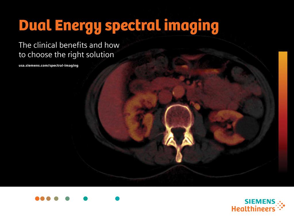
Discover Dual Energy (DE) spectral imaging. It’s the difference between images and answers. Visualization and characterization. Qualification and quantification. Built into all of our CT scanners, it delivers powerful performance, incredible versatility for your entire patient population, and exceptional ease-of-use – all while integrating seamlessly with your current workflow.

And that is a world of difference.

Features & Benefits

Dual Energy examinations provide answers for a wide range of diagnostic challenges. With Siemens SOMATOM® CT scanners, you receive excellent Dual Energy images at equal, or often lower dose levels compared to conventional CT examinations. This makes Dual Energy a valid modality for many types of patients and a wide variety of clinical questions. That’s what True Dual Energy is about.


DEfinitely excellent images

Dual Energy from Siemens Healthineers – True Dual Energy – offers crisp image quality with no extra radiation dose. In addition, Dual Energy CT offers you clinical information beyond morphology and allows you to highlight, characterize, quantify, and differentiate the material in your scans. Here are three examples:

Optimum Contrast for sharper image contrast

Optimum Contrast for sharper image contrast
Optimum Contrast is an intelligent method that changes the blending ratio of low and high energy data on a pixel-by-pixel basis, depending on the corresponding Hounsfield units (CT numbers). For higher CT numbers, which occur in regions of high iodine concentration, larger proportions of the image are taken from low tube energy data. As a result, areas of contrast enhancement are accentuated.

Monoenergetic imaging for reducing metal artifacts

Monoenergetic imaging for reducing metal artifacts
With Monoenergetic imaging, metal artifacts can be reduced in both Single Source and Dual Source Dual Energy data. Select the energy level at which implants, clamps, or screws will have the smallest impact on image quality and get unsurpassed scan results.

Electron density and effective atomic number maps in one examination
The attenuation of X-ray, next to others, depends on the electron density and the effective atomic number. Both parameters are characteristic for different materials. Thus syngo.CT DE Rho/Z may aid in the differentiation and characterization of different tissues in the body.


DEfinitely the right dose

Only with Siemens Healthineers' SOMATOM CT scanners, you get all advantages of True Dual Energy imaging with no dose penalty in either Single Source or Dual Source Dual Energy scans. With the latest dose reduction techniques, such as the Tin Filter, ADMIRE, and CARE Dose4D, you will find the right dose level for generating valuable diagnostic information at the best possible image quality.

Following the ALARA principle, Siemens Healthineers has introduced Dual Energy scan modes that utilize all available dose reduction techniques – such as CARE Dose4D™ for real-time mA modulation and SAFIRE/ADMIRE, to reduce the mA created by iterative reconstruction.

Dose reduction in Dual Spiral Dual Energy
In the Dual Spiral Dual Energy scan mode, two consecutive spiral scans are performed at approximately one-half the dose so that you can be certain of compliance with the ALARA principle.

Dose reduction in TwinBeam Dual Energy
TwinBeam Dual Energy consist of a regular 120 kV spectrum filtered by different material. As it is a normal spiral scan all dose reduction features can be applied, such as Care Dose4D and/or SAFIRE¹ /ADMIRE². The TwinBeam Dual Energy technique does not have any dose penalty, thus it always follows the ALARA principle. 

CT DE Booklet Graphic
The Tin Filters enables dose dose neutrality by eliminating spectral overlap

Dose reduction in Dual Source Dual Energy
SOMATOM Force and SOMATOM Drive with its Tin Filters further established Dual Energy as a dose-neutral technology. The Tin Filters filters out unnecessary photons from the high-energy X-ray tube. It permits the use of Dual Energy in daily practice at a dose comparable to a conventional 120-kV scan, with optimal spectra separation for excellent Dual Energy data quality.


Dual Energy for all patients

Every patient is different. But True Dual Energy offers you dedicated protocols and evaluation software applications for virtually all patients and a wide range of clinical questions.

Even for difficult patient conditions
You can use Siemens Healthineers’ Dual Energy with all Single Source CTs ranging from SOMATOM Scope Power, SOMATOM Perspective, and SOMATOM Definition AS family to SOMATOM Definition Edge. In addition, with SOMATOM Definition Flash, SOMATOM Drive, and SOMATOM Force you get the outstanding Dual Source CT.

Compliance with the ALARA principle allows you to tap the advantages of Dual Energy with a very wide range of patients – including children as well as older, obese and fragile patients. On top of that, optimized scan protocols let you scan at low contrast agent dose and flow rate.

Wide range of applications for fast, definitive answers
Thanks to an ever-growing spectrum of applications, True Dual Energy helps you find fast and confident answers to a wide range of clinical questions – from lung nodes to brain hemorrhage, from perfusion defects in the heart to the evaluation of the malignancy of tumor lesions.


Clinical Use

Siemens Healthineers' Single Source and Dual Source Dual Energy techniques impress radiologists with their easy-to-use scan protocols, their suitability for daily clinical routine, their range of applications, and their scientific validation (more than 200 scientific publications).


Brain Hemorrhage

Differentiate hemorrhage from iodine-uptaking bleedings and lesions
The Dual Energy application syngo.CT DE Brain Hemorrhage assists you in visualizing iodine concentration and distribution in the brain. Lesions and bleedings may show significant iodine uptake in the image, while inactive hemorrhages are not enhanced.


syngo.CT DE Gout

Accurate and non-invasive diagnosis of gout
The diagnosis of gout is not always simple – for example, in acute cases where an aspiration cannot be performed. Nor is the uric acid concentration in the blood always a reliable indicator. syngo.CT DE Gout overcomes these limitations by quickly, noninvasively, and accurately visualizing uric acid crystals. This spares your patient a painful and stressful examination.


syngo.CT DE Heart PBV

Reveal perfusion defects with only one scan
With syngo.CT DE Heart PBV, you can visualize the iodine concentration and distribution in the myocardium to reveal perfusion defects. In addition, you can use a virtual non-contrast display to identify myocardial edema. The simultaneous acquisition of high- and low-kV datasets diminishes the problem of misregistration due to cardiac motion.


syngo.CT DE Calculi Characterization

Characterization of kidney stones
Kidney stones contain varying amounts of heavy atoms (for example, uric acid stones versus calcified stones). Dual Energy CT can be used to perform a differentiation of these materials. With syngo.CT DE Calculi Characterization, you can visualize these chemical differences in kidney stones and use the information for your therapy decision.


syngo.CT DE Direct Angio

Accurate bone removal even in complex body regions
With syngo.CT DE Direct Angio, you can overcome the limitations of conventional bone removal software. The Dual Energy approach reliably isolates even complex vasculature, for example, at the base of the skull where CTAs are difficult to interpret.


syngo.CT DE Virtual Unenhanced

Quantify lesions with two scans in one
syngo.CT DE Virtual Unenhanced assists you in visualizing the contrast agent concentration in the liver. The basis for this approach is a material decomposition into iodine contrast agent, fat, and liver tissue. You can also create virtual non-contrast information using the Dual Energy information from fat and liver material. You can switch between these two representations of the anatomical information without the need for an additional non-contrast scan.


syngo.CT DE Lung Analysis

Assessment of perfusion defects and affected vessels at a glance
Perfusion defects in the lung are often caused by pulmonary embolism. The application syngo.CT DE Lung Analysis enables the color-coding of vessels that are affected, for example, by pulmonary emboli. It also allows a fast evaluation of lung perfusion defects at a glance without requiring an additional non-contrast scan.


Monoenergetic imaging

Customize your image impression – reducing metal artifacts
Metal artifacts disturb your diagnostic image quality and your therapy planning. With syngo.CT Dual Energy’s Monoenergetic images, you can reduce metal artifacts to get the results you need by selecting the energy level with the best contrast-to-noise ratio and a minimum of metal artifacts.


Dual Energy Mono

Maximize image quality with new algorithm
Allows users to display monoenergetic images for a range of 40–190 keV. By displaying multiple monoenergetic ROIs and the associated absorption curves, Monoenergetic Plus lets users easily compare and quantify lesions and tissues. The ability to export statistical information into the file system for further evaluation is very beneficial for various research and diagnostic tasks.

Our new video presented by Prof. Wichmann and his team from the University Hospital Frankfurt shows the clinical value of Dual Energy by using Monoenergetic Plus images. The video explains how Rapid Results speeds up your post-processing workflow with our Zero Click Solution.


ct Dual Energy Bone Marrow

Characterizing different materials
The attanuation of X-ray, next to others, depends on the electron density and the effective atomic number. Both parameters are characteristic for different materials. With syngo.CT DE Rho/Z you have access to electron density and effective Z maps in one examination.


Bone Marrow

Evaluation of the bone marrow
Bone marrow can be affected by various pathologies, such as bone bruises after trauma and diffuse tumor infiltrations.
syngo.CT DE Bone Marrow allows for the segmentation and visualization (color-coding) of the bone marrow based on a material decomposition into bone marrow and calcium.


Computed Tomography Dual Energy Hardplaque Cut

Differentiate calcified plaques from iodine contrast
syngo.CT DE Harplaque Display enables the identification (color-coding) and automatic removal of calcifications from a Dual Energy CTA image. By therefore differentiating between hard plaques and contrast agent this Dual Energy application helps to display the true vessel lumen without interfering hard plaques.

Technical Specifications

Dual Energy has opened the door to a new dimension of CT diagnosis: the characterization and visualization of chemical composition is no longer in the realm of science fiction. As the creators of the world’s first Dual Source Dual Energy CTs, we have acquired a lot of experience – and we have drawn on that experience to design our Single Source Dual Energy scanners as well. Here’s how our technologies work:


DE Single Source
The dose-optimized Single Source Dual Energy scan mode consists of two consecutive scans at different kV values, each at one-half the dose.

The Dual Spiral Dual Energy scan mode consists of two successive spiral scans at different kV and mA levels. It is available for SOMATOM® Definition Edge, SOMATOM Definition AS family, SOMATOM Perspective, and SOMATOM Scope Power – you can do more than just morphology with Single Source CT.


Software modules available for Dual Spiral Dual Energy*

The easy-to-use scan mode makes Single Source Dual Energy available for your daily routine 24/7. Dual Spiral Dual Energy comes with dedicated applications for your clinical area. In addition, it features the syngo.CT Dual Energy1 basic viewer with Optimum Contrast and monoenergetic imaging.


  • syngo.CT Dual Energy1 – basic viewer including syngo.CT DE Rho/Z and monoenergetic imaging
  • syngo.CT DE Calculi Characterization1
  • syngo.CT DE Gout1
  • syngo.CT DE Monoenergetic Plus1 
  • syngo.CT DE Bone Marrow1
  • syngo.CT DE Virtual Unenhanced - Liver VNC1
  • syngo.CT DE Brain Hemorrhage1 


TwinBeam Dual Energy

This innovative Dual Energy approach based on a split filter at the X-ray tube allows simultaneous acquisition of high and low kV datasets in single source systems, enabling all high contrast dynamic applications. TwinBeam is available for SOMATOM Definition Edge and SOMATOM Definition AS+.

Software modules available for TwinBeam Dual Energy

TwinBeam Dual Energy enables simultaneous acquisition of high and low kV datasets. Therefore it is possible to use all available True Dual Energy applications on Single Source CT systems with no compromise on dose.


  • syngo.CT Dual Energy1 – basic viewer including Optimum Contrast, syngo.CT DE Rho/Z and monoenergetic imaging
  • syngo.CT DE Virtual Unenhanced1
  • syngo.CT DE Brain Hemorrhage1
  • syngo.CT DE Direct Angio1
  • syngo.CT DE Lung Analysis1
  • syngo.CT DE Calculi Characterization1
  • syngo.CT DE Gout1
  • syngo.CT DE Hardplaque Display1
  • syngo.CT DE Monoenergetic Plus1


DE Dual Source
The Selective Photon Shield ensures dose neutrality by eliminating spectral overlap. This makes Dual Energy as dose-efficient as any single 120 kV scan.

During a Dual Source Dual Energy scan, two CT datasets are acquired simultaneously with different kV and mA levels, allowing you to visualize differences in the energy-dependence of the attenuation coefficients of different materials. These images are combined and analyzed to visualize information about anatomical and pathological structures. Dual Source Dual Energy scanning is available for SOMATOM Definition Flash, SOMATOM Drive, and SOMATOM Force.


Software modules available for Dual Source Dual Energy

Dual Source Dual Energy comes with dedicated applications for your clinical area. In addition, it features the syngo.CT Dual Energy1 basic viewer with Optimum Contrast and monoenergetic imaging. All of these modules facilitate sound and sustainable treatment decisions.


  • syngo.CT Dual Energy1 – basic viewer including Optimum Contrast, syngo.CT DE Rho/Z and monoenergetic imaging
  • syngo.CT DE Virtual Unenhanced1
  • syngo.CT DE Brain Hemorrhage1
  • syngo.CT DE Direct Angio1
  • syngo.CT DE Lung Analysis1
  • syngo.CT DE Heart PBV1
  • syngo.CT DE Calculi Characterization1
  • syngo.CT DE Gout1
  • syngo.CT DE Hardplaque Display1
  • syngo.CT DE Bone Marrow1
  • syngo.CT DE Monoenergetic Plus1


1