At IDKD Asia 2025 in Singapore, one lecture drew attention for its focus on clinical practice and practical insights. In her session titled “Workstations as the New Cornerstone of Thoracic CT Imaging,” Dr. Lynette Teo from National University Hospital Singapore, issued a clear message: The future of radiology hinges on the intelligent workstation.
“It’s not just where we read images. It’s where we extract value,” she said with conviction, pausing to let the message land.
Dr. Teo’s talk, combined with technical insights and broader perspective, outlined the progression of radiology from film to AI, from static images to quantitative analysis, and ultimately from passive viewing to precision medicine powered by workstations.
From Pixel to Precision: The New Role of Workstations
Modern thoracic CT demands more than detection - it demands context. Today’s workstations offer:
- Nodule detection and volumetry to support Low Dose CT (LDCT) screening
- Cavity analysis in TB and infections
- Quantitative fibrosis and emphysema scoring using Hounsfield density maps
- Pulmonary perfusion mapping through dual-energy CT
- 3D vascular reconstructions and airway evaluations
She shared multiple case examples including a post-lobectomy patient with suspicious nodules and a systemic sclerosis case with subtle Interstitial Lung Disease progression - highlighting how AI-powered volumetry and CT densitometry aided confident diagnosis and follow-up.
Lung Cancer in Singapore: A Call for Screening - and Better Tools
Dr. Teo provided local context by presenting these statistics: Lung cancer remains the top cause of cancer deaths in Singapore, and nearly half of non-small cell lung cancer cases are in never-smokers. Yet no national screening program exists.1
“LDCT gives us eyes, but workstations give us clarity ,” she said, pointing to how CAD tools reduce oversight and support longitudinal follow-up with reproducible quantification.
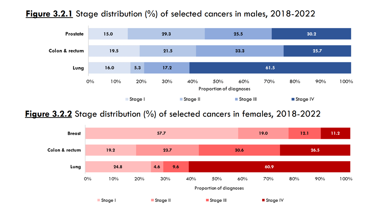
Singapore Cancer Registry Annual Report 2022, National Registry of Diseases Office, Sept 20242
Why Radiologists Must Embrace the Workstation
Why Radiologists Must Embrace the Workstation
- Radiologist workloads are skyrocketing, but workstation tools can speed up quantification.
- Structured reporting integrated with measurements can reduce error and streamline MDT discussions.
- AI tools, when used judiciously, augment judgment—not replace it.
She emphasized the need to practice with the workstation, echoing her early experience with Siemens Leonardo and now with syngo.via VB60/VB80 platforms.
“Workstations are only as good as the hands that use them. Having dedicated over two decades and more than 10,000 hours to advanced post-processing at the Siemens workstation, I’ve witnessed firsthand the platform’s transformative impact on diagnostic imaging. I'm eager to harness the capabilities of the next-generation syngo.Via, now enhanced by AI-driven workflows and quantitative analytics.”
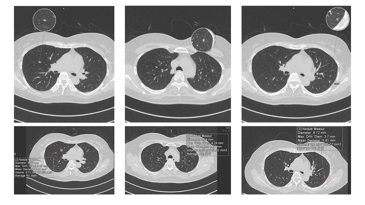
Image courtesy from National University Hospital, Singapore illustrating computer assisted diagnosis (CAD) of lung nodule on syngo.via VB60
The AI Companion: Friend or Foe?
She demonstrated the Siemens Healthineers’ AI-Rad Companion Chest CT, showcasing its segmentation of nodules, lung parenchyma, fibrosis, and even the aorta. But she also cautioned:
- “Bias in training data still exists.”
- “Not all findings flagged are real—AI has a learning curve, just like we do.”
- “Use it to confirm, not to diagnose blindly.”
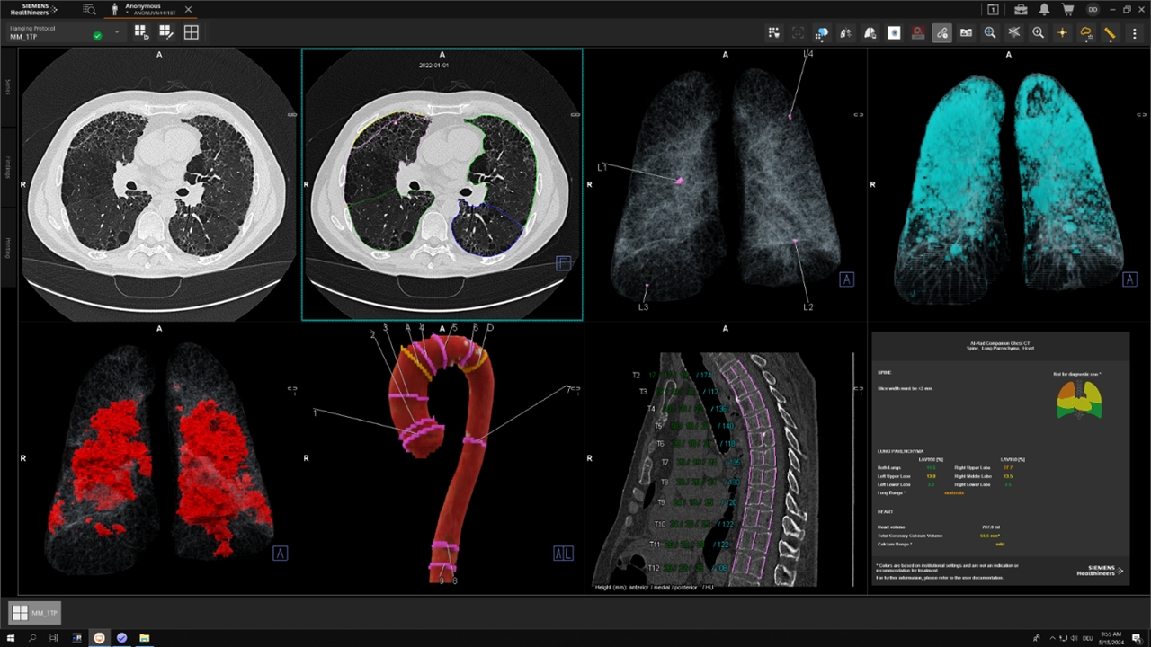
The AI-Rad Companion Chest CT detects and highlights lung nodules.
Photon-counting and the Road Ahead
In this part of her talk, Dr. Teo introduced photon-counting CT as the next leap in transforming medical imaging. With submillimeter resolution and reduced radiation, scanners like the NAEOTOM Alpha enable reclassification of ILDs and lower-dose follow-ups.
“You can now see what used to be a blur. But if your post-processing can’t keep up, it’s like watching HD on a broken screen.”
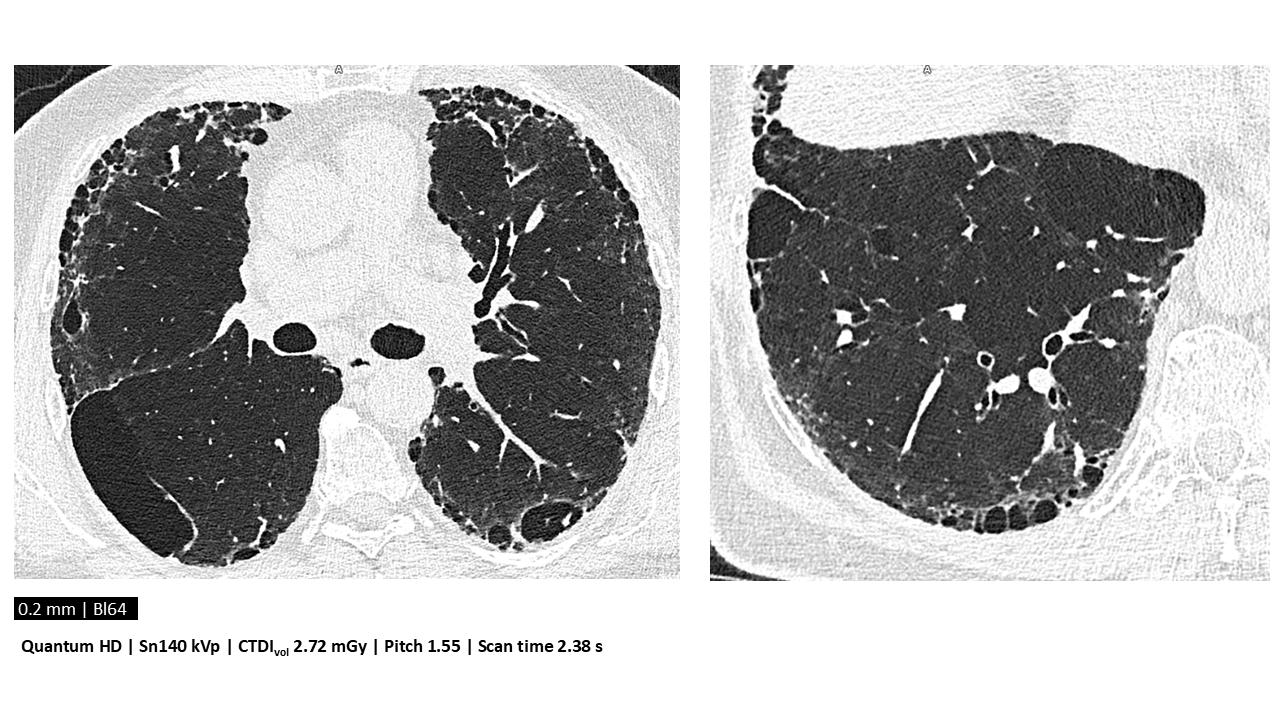
Lung assessment combining Quantum HD images with low radiation dose
Courtesy of Erasmus Medical Center Rotterdam, Rotterdam, The Netherlands
Take-Home Messages
Her final message:
“The workstation is not just a machine. It’s our clinical ally, our second pair of eyes, our quantifier of uncertainty. Invest the time to master it—because precision medicine begins at the workstation.”
12th IDKD Asia 2025: Disease of the Chest, Heart and Vascular System, May 29 – 31, 2025, Singapore
This presentation “Workstations as the New Cornerstone of Thoracic CT Imaging,” was presented by Dr. Lynette Teo from National University Hospital Singapore
IDKD Asia 2025 in collaboration with National University Hospital Singapore provide interactive education for medical imaging specialist and others interested For more information, click this link: https://idkd.org/cms/home
Written by

Dr Lynette Teo
is a Senior Consultant Radiologist at the National University Hospital, Singapore, and holds Associate Professorships at both the National University of Singapore and Vin University, Vietnam. She completed her Radiology training in Singapore before advancing her expertise with cardiothoracic imaging fellowships at the Royal Brompton Hospital in the United Kingdom.
With over a decade of experience as a program director, Lynette remains deeply committed to shaping the future of radiology education. She plays a role in undergraduate and postgraduate training, serving as an examiner for the conjoint Singapore-Royal College of Radiologists examination, an accreditor for the Joint Committee of Specialist Training in Singapore, and deputy chair of the local ethics board. She actively contributes to several national health committees. Lynette's research interests centre on artificial intelligence applications in cardiac CT, leading to the co-founding of an AI start-up. She is also interested in harnessing 4D flow CMR for congenital heart disease. She is involved in several multicentre cardiothoracic CT and MRI imaging trials and has published more than 120 articles in peer-reviewed journals.