High-V MRI – Value beyond barriers
Digitalization is rapidly transforming MR imaging by applying highly efficient acquisition techniques and deep learning-based reconstruction. High-V MRI takes the power of digitalization and deliberately applies it to a new field strength of 0.55T with inherent clinical benefits. High-V MRI combines the best of both worlds to offer a new era in MRI that embraces diagnostic confidence in daily routine and new clinical opportunities.
The digital transformation of MRI
Siemens Healthineers is bringing the digital revolution to your MRI practice. We are constantly perfecting our acquisition technologies to achieve higher image quality while reducing patient slot times. And now with Deep Resolve, we have introduced deep learning-based reconstruction to further enhance the quality of outcomes. MAGNETOM Free.Max leverages the full potential of the digital power of Siemens Healthineers for the utmost in diagnostic quality.

Deep Resolve Gain
Deep Resolve Gain is an intelligent algorithm that uses individual quantitative noise maps in the reconstruction process for targeted denoising. It is available for a broad range of sequences and boosts image quality for maximum diagnostic capabilities.


Image-ID: 1aaaa3623 | 4aaaa0399
Improved implant imaging with High-V MRI
Imaging metal implants has historically been difficult with conventional MRI systems as metal causes artifacts. High-V MRI offers intrinsic physical advantages that result in reduced metal distortions and therewith strongly improved diagnostic capabilities for implant imaging.

Image Courtesy: University Hospital Erlangen, Germany | Image-ID: 4aaaa0419
New opportunities in pulmonary imaging with High-V MRI
Pulmonary imaging has been notoriously difficult with conventional MRI as the air-tissue interfaces result in a fast signal decay. These challenges scale with magnetic field strength which makes High-V MRI the perfect opportunity for pulmonary imaging. As a result, High-V MRI provides possibilities to expand the reach of pulmonary MRI.
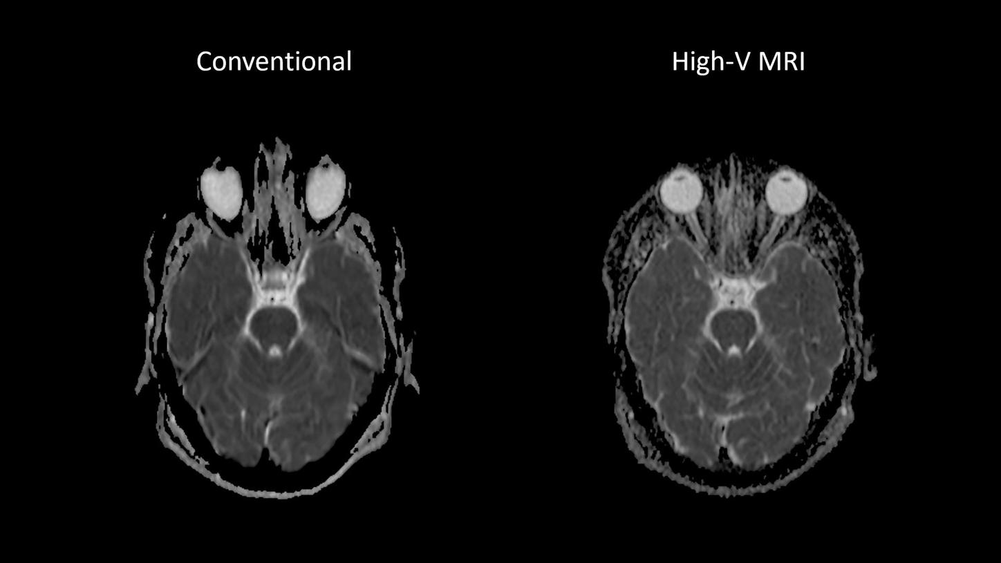
Image-ID: 1aaaa3439 | 4aaaa0297
Reduced susceptibility challenges with High-V MRI
Susceptibility artifacts are a well know phenomena in MRI. One prominent example for such artifacts are at air-tissue interfaces, as they occur at the sinuses and orbits. The unique field strength of High-V MRI offers physical advantages that inherently reduce susceptibility artifacts. This leads to reduced geometric distortions in diffusion imaging which results in improved diagnostic quality.
Did this information help you?
The information shown herein refers to products of 3rd party manufacturer’s and thus are in their regulatory responsibility. Please contact the 3rd party manufacturer for further information.
The product is still under development and not commercially available yet. Its future availability cannot be ensured.