Addressing the rising prevalence of Non-alcoholic Fatty Liver Disease
Learn more about NAFLD, a leading cause of liver related mortality
Non-alcoholic fatty liver disease (NAFLD) is a condition in which excess fat, not caused by heavy alcohol use, is stored in the liver. There are two types of NAFLD: simple fatty liver (simple steatosis) and non-alcoholic steatohepatitis (NASH).
NAFLD is projected to become the leading cause of liver-related mortality within 20 years.1
Infographic: Causes of liver diseases
Several factors can lead to liver disease. Excessive alcohol consumption, obesity, diabetes, hepatitis infections, and excessive consumption of medication could all contribute to an inflamed, and eventually fibrotic, liver.

Liver damage

Normal liver
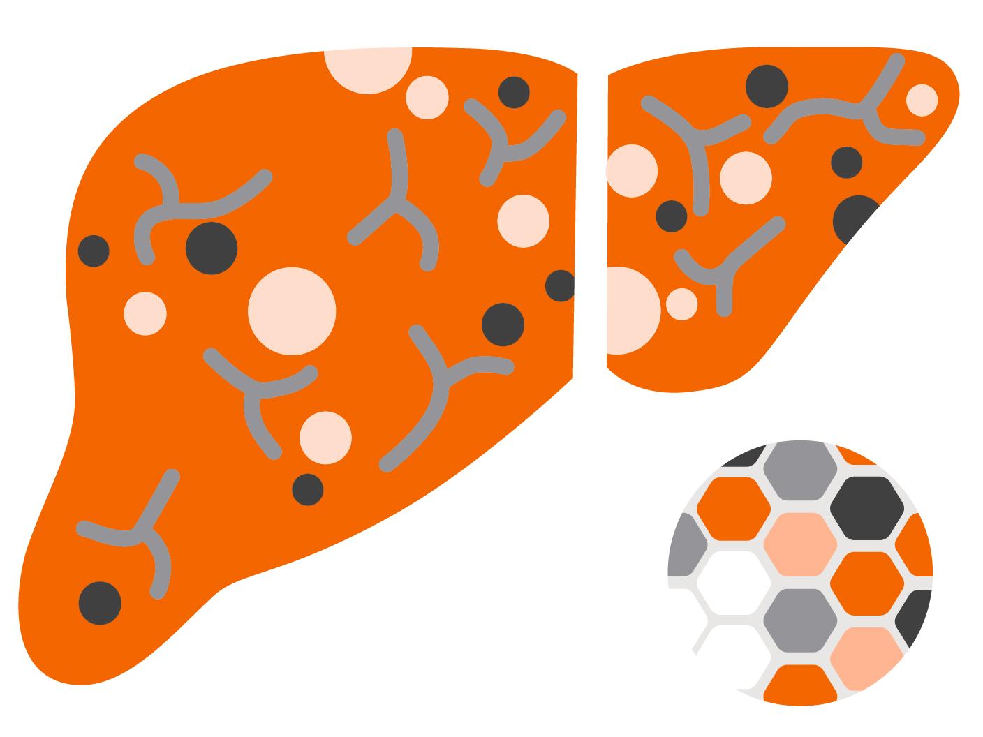
Lobular structure of the liver
The liver contains an estimated 1 to 1.5 million hepatic lobules with a diameter of 1-2mm.
Hepatic lobules are small structural units composed of liver cells (hepatocytes).

Inflamed liver
Formation of collagenous connective tissue
If liver cells are chronically damaged—for example, by a prolonged inflammation—excessive collagenous connective tissue accumulates.
Fibrotic liver
Hardening of the liver
The connective tissue gradually replaces the actual liver cells. The organ becomes scarred and loses its elasticity and function.
Progression of liver disease
Click on each item to learn more about the stages and progression of liver disease.


How liver assessment works
Click on the cards below to learn about different types of liver assessment.

Elastography
A probe emits a mechanical pulse toward the liver.

An integrated ultrasound transducer measures the velocity of the pulse wave between two points. The less elastic the liver tissue, the faster the pulse propagates through the liver.

Biopsy
A tissue sample is taken from the liver with a cannula.

Biopsy
The sample is then examined for scar tissue under a microscope.

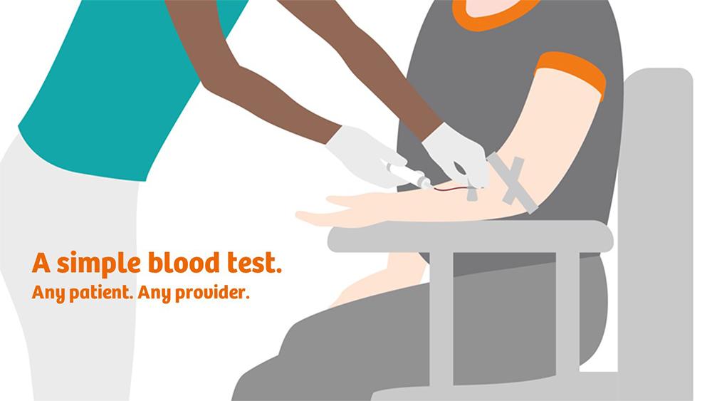
The ELF Test
A blood sample is taken.

The ELF Test
Three important serum markers can be detected with an automated analyzer and the risk of disease progression can be derived from these.
Current challenges in NAFLD patients
Among the current and growing number of NAFLD patients, there is an urgent need for the early and accurate identification of patients at risk of progressing to cirrhosis and liver-related events (LRE). Patients with mild disease are often inappropriately referred to secondary care for invasive investigations and undiagnosed patients remain in primary care until complications of cirrhosis develop.
Click below to learn more about challenges in NAFLD patients.
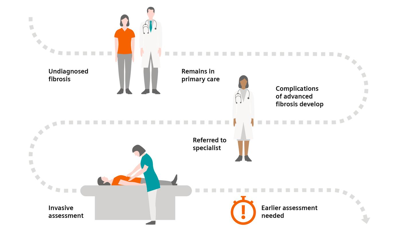
The need for non-invasive Liver Fibrosis tests
Assessment of liver fibrosis has traditionally relied on costly and invasive liver biopsy that requires a specialist, may not be representative of the amount of fibrosis, and carries a risk of life-threatening complications. Follow each step of the pathway from undiagnosed fibrosis to invasive assessment.
Did this information help you?
The products/features (mentioned herein) are not commercially available in all countries. Their future availability cannot be guaranteed.
Younossi ZM, et al. Hepatology. 2016;64:73–84.
Pais R, Barritt AS 4th, Calmus Y, et al. J Hepatol. 016;65(6):1245–1257. doi:10.1016/j.jhep.2016.07.033.
Younossi ZM, et al. Diagnostic modalities for nonalcoholic fatty liver disease, nonalcoholic steatohepatitis, and associated fibrosis. Hepatology. 2018 Jul;68(1):349-60.
Anstee QM, et al. Noninvasive tests accurately identify advanced fibrosis due to NASH: baseline data from the STELLAR trials. Hepatology. 2019 Nov;70(5):1521-30.