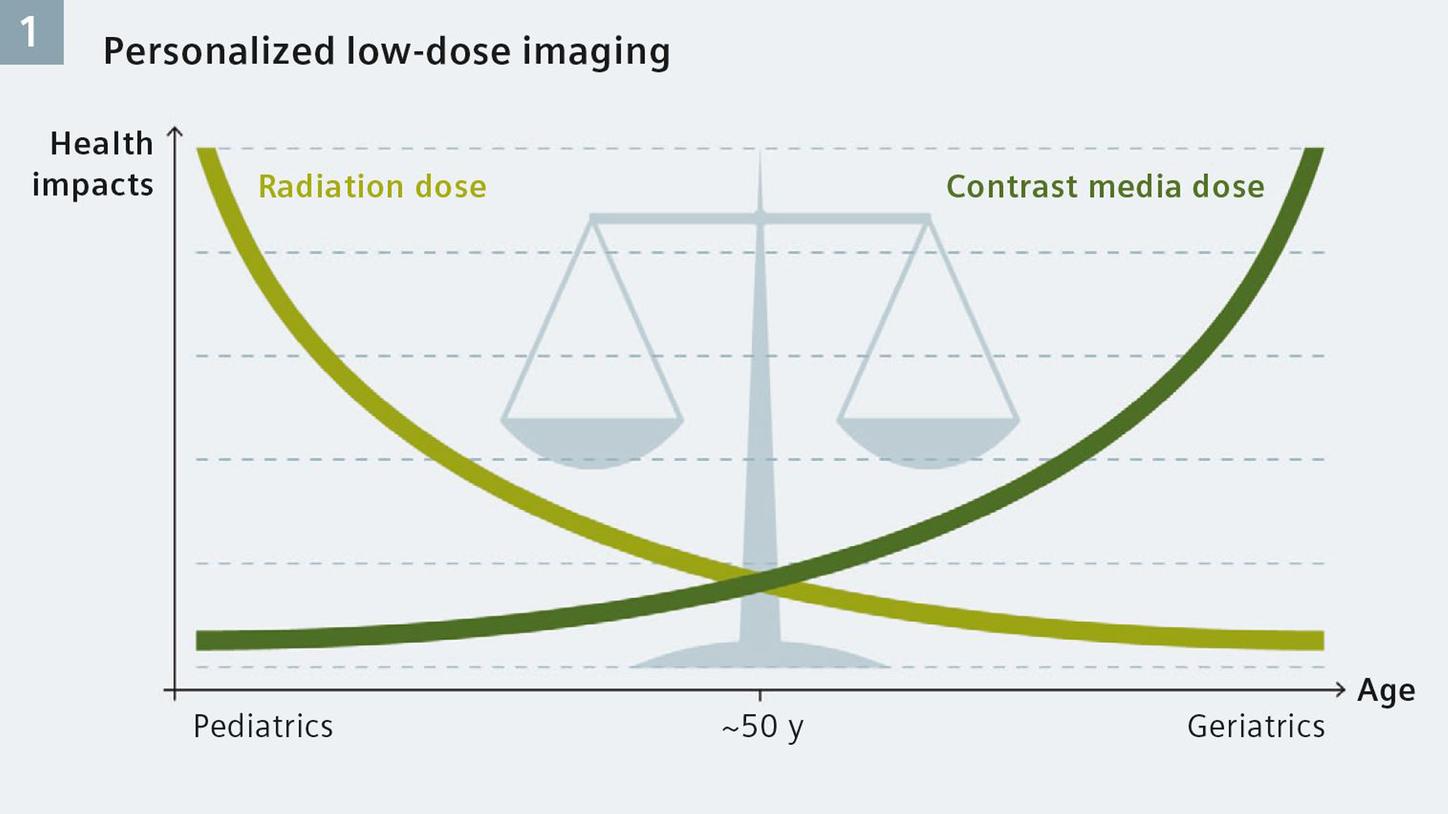
Reducing doses of radiation or contrast media has a significant impact on all patients.
With this in mind, physicians, researchers, and providers strive constantly to apply these two critical components in CT scanning, X-ray and contrast media, in doses that are as high as necessary and as low as possible.
Saving iodine by lowering tube voltage
Tube potentials between 70 and 100 kVp have shown to be most effective for clinical contrast CT examinations. The basis for low kV imaging is the mass attenuation coefficient, which is a property that depends on the chemical composition and density of a material. Early clinical experience based on imaging of the left ventricle and aortic root (TAVI studies) demonstrate that a reduction in contrast media administration may be possible using SOMATOM Force’s Turbo Flash Mode and its low kV/high mA capabilities.
Phantom scans show a linear relationship between the iodine concentration and enhancement, and how much iodine can be saved if the attenuation is kept constant. For example: Compared with a 120 kV baseline protocol, the iodine enhancement at 100 kV is higher and therefore the contrast can be reduced by 20 mL if the original volume was 100 mL (For all other values, please see the dedicated white paper). The principle works on every CT scanner.
However, meaningful savings of contrast media dose require a significant decrease in kV. An animal study in pigs demonstrated that an identical temporal enhancement curve for the 120 kV reference protocol (Fig. 2A) and 70 kV protocol (Fig. 2B) can be obtained with constant injection duration and adapted iodine delivery rate (IDR) while reducing the contrast media from 64 mL to 32 mL (see Fig. 2).[1]
Adapted contrast injection protocol for reduced iodine load
To reduce contrast agent dose and change from a reference scan protocol to a low kV protocol, the contrast injection protocol must be adapted to reduce the iodine load. The easiest way to adapt the iodine load would be to reduce the contrast volume without changing any other injection parameters. However, a reduction in contrast volume leads to shorter injection duration and, therefore, to significant changes in contrast enhancement over time: Peak enhancement occurs earlier, requiring adaptation of the scan delay, and scan timing becomes more critical because of a narrower enhancement curve.
Various institutions pursue the concept of low kV scanning. Researchers from the University of Frankfurt have published initial clinical results from 70 kV ultra-low dose CT scans of the paranasal sinus.[2] Researchers at University Hospital Mannheim reduced the amount of contrast agent for dedicated angiographic CT examinations to 20 mL for a diagnostic CT angiography of the chest, abdomen, and pelvis.
To find out more about the use of contrast enhancement at lower kV to reduce the contrast agent dose while maintaining high image quality in terms of contrast-to-noise (CNR), please follow the link below to the related full-length white paper.
About the Author
Ivo Driesser, Siemens Healthcare, Germany

![A preclinical animal study demonstrated the peak width, peak height and peak time remain the same with a reduction in the iodine delivery rate.[1]](https://marketing.webassets.siemens-healthineers.com/1800000005412952/83d01718cb9b/v/13dd6e8791f2/Scanning_at_low_kv_Image-2a-b_Neu_1800000005412952.jpg)