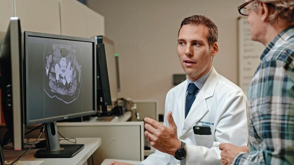
With more information comes more complexity. That is why radiologist Daniele Marin, MD, at Duke University Medical Center welcomes any support that can make CT scanning easier, faster, and more reliable.
Download your print version here.
Photos: D. L. Anderson
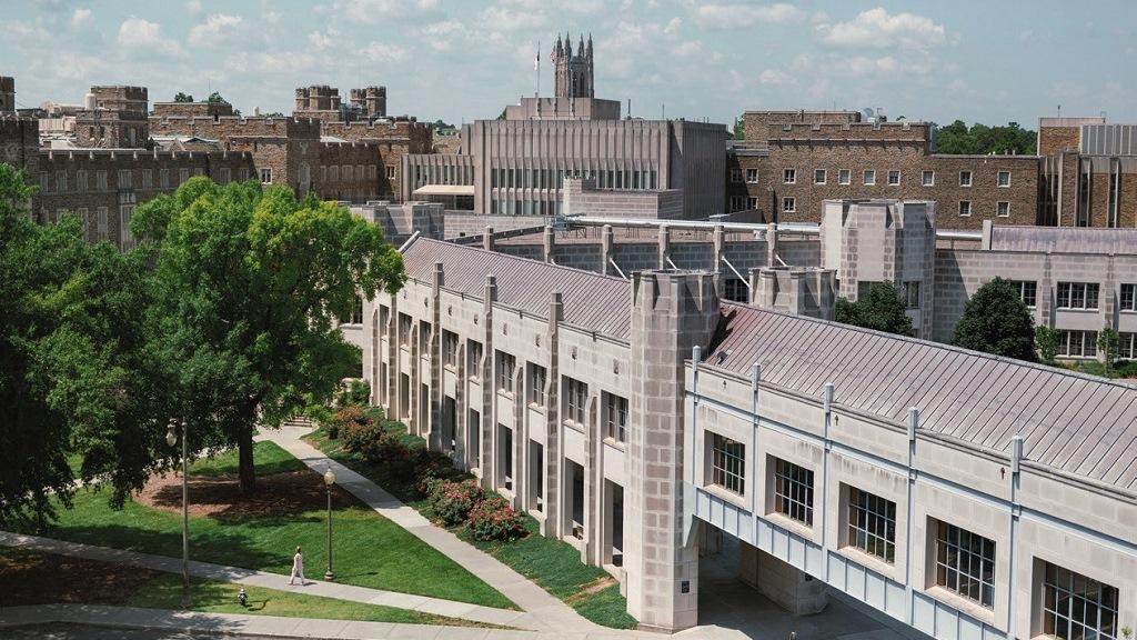
"Sapienza” is Italian for wisdom. It is also the name of the world-famous university in Rome where Daniele Marin, MD, took the first steps in his academic career. His quest for medical wisdom led Marin to Duke University Medical Center, where today he is Associate Professor of Radiology. Located in Durham, North Carolina, Duke University is one of the cornerstones of the famed “Research Triangle”. Its clinics offer top-notch medical treatment and medical research in all relevant fields. “Duke offered exactly the research opportunities I was looking for,” says Marin, who came here from Italy ten years ago. On the sprawling campus of the Medical Center, new research and treatment facilities are being built regularly. The 50 faculty members of Duke’s Radiology Department oversee roughly 20 computer tomography platforms, “Clinically we are a very busy department.”
Radiology is based on perception
For Daniele Marin this translates to a large number of case readings every day. “Of course we have residents and fellows, so it’s not like it’s only me sitting at a workstation,” he says. “Five team members are in the room. Each has three to four patients, and I hop from place to place. Usually I read between 50 and 70 cases a day.” However, the radiologist is never overwhelmed, “I love anatomy,” he says. “It has always been my passion to see into the patient, to see structures and organs. That’s why I fell in love with radiology.” As Director of the Multi-Dimensional Image Processing Lab Marin finds the capabilities of Dual Energy CT uniquely productive and has realized some of the benefits of this sophisticated technology. The lab is located at the Hock Plaza Building in the western side of the campus and employs more than half a dozen highly specialized technologists. In subdued lighting, they do sophisticated image postprocessing work at workstations, each equipped with three flat-screen monitors. Acquiring experience is indispensable for Marin’s professional growth. “My ultimate goal is to advance CT imaging,” he says, “Radiology is very subjective, based on a perception of the radiologist. And a lot of the decisionmaking is based on this perception. My goal is that in the future we can provide more meaningful, more quantitative, more measurable metrics of patient response to therapies, including predictions of outcomes.”

Full spectrum of details
At Duke, Dual Energy CT scans are routinely done in four areas: Neural scans help to map brain hemorrhages; chest scans are used to locate pulmonary embolisms and create perfusion maps; musculoskeletal scans visualize uric acid crystals that my indicate gout and pathologies of bone marrow. Daniele Marin is most interested in the abdominal area, where dual energy is very powerful in analyzing lesions of the liver and kidney. As an example, Marin shows the dual energy image of a kidney. “A lower monoenergetic level is better for visualizing lesions, for example diffuse tumors, because it enhances the attenuation and therefore its conspicuity,” he explains. The radiologist points to a very small bright spot. With dual energy I have the full spectrum of energies at hand. I can choose the level that best visualizes what I need. For instance, a high monoenergetic level is very useful in reducing metal artifacts. Moreover, iodine uptake and a virtual noncontrast scan are also calculated, which helps characterize and measure a lesion very accurately.
Marin remembers a case when dual energy was crucial in a time-critical situation. A patient in the emergency department was suffering from intestinal bleeding. Before we had implemented dual energy routinely, the technologist was supposed to do a CT scan without and then with contrast media, but he forgot the first round. Luckily the scan was done with dual energy and the radiologist was still able to calculate a virtual noncontrast and differentiate low density material from high density materials, which may be a sign of bleeding in the bowel. “We were able to rule out a source of the bleeding,” Marin says. “Dual energy can help a lot avoiding such mistakes.”
“I think it’s critical to make information available to more of the people who need to see it.”
Automation is cost-effective
Dual Energy CT is also cost-effective. For example, a patient comes into an emergency room with bowel inflammation. The physicians suspect diverticulitis and perform an abdominal CT scan. On the images they don’t see diverticulitis, but rather a lesion in the kidney. The scan was performed with dual energy thus the images allow for a preliminary diagnosis. Immediate follow-up scans are unnecessary. For Daniele Marin, this hypothetical case illustrates one of the benefits of Dual Energy CT; it can reveal more incidental findings. “It has the potential to decrease the overall financial burden for the health system.”
Duke’s radiologists fully embrace the concept. Accordingly, “dual energy is becoming a large proportion of what we are doing,” says Marin. He estimates that the department conducts up to 500 dual energy scans per month. But even a top-of-the-line research institution, such as Duke, sometimes struggles with the complexity and additional time requirements that come with this process. “Workflow is one of the most important challenges,” Marin says. At Duke we were typically performing two reconstructions for single energy scans in the abdominal field-one on the axial plane, and one coronal. “With dual energy, there is the opportunity to reconstruct a lot of additional series, with the subsequent extra information. We move to five, eight different datasets for the same scan.”

Without a fitting workflow solution, the increase in time required is substantial. Radiologists report that the reconstruction of a dual energy scan takes 20 minutes as opposed to 5 minutes for single energy. If technologists do all the extra postprocessing work at the CT console, the number of reconstruction mistakes could increase. Marin estimates a 5-10 percent chance of error. These drawbacks largely disappear with Siemens Healthineers’ Rapid Results Technology. In its new iteration, this software solution enabled by the syngo.via reading and reporting platform automates all of the complicated postscanning calculations. “The original datasets are sent to the syngo.via server,” Marin explains, “This computer automatically reconstructs all the different series I need. You minimize the time at the CT console, and you minimize the chance of reconstruction mistakes. You are automating the process.”
Providing a faster workflow
Siemens Healthineers’ most advanced dual energy capable scanner, SOMATOM Force, is located in the newly erected Oncology Center. The scanning process itself does not take longer when done with dual energy. “Acquiring the images pretty much requires the same amount of time as with single energy,” says Daniele Marin. After the patient passes through the scanner gantry, which is now glowing in pleasant colors, the technologist at the console finishes the image acquisition and the data is automatically sent to be processed. Within a few minutes he is ready to accept the next patient. While the new patient is positioned on the scanning table, the off-site computers generate preprogrammed datasets and format the resulting images to be evaluated by the radiologist in the PACS system. “It is very important that all of these series are available on PACS,” Marin says, “since it is everybody’s natural reading environment.” In this way, Rapid Results Technology makes information available to more of the people who need to see it. “I think it’s critical.”
Information can be streamlined
Experience tells the radiologists that automation tools will not hide information that a manual process would reveal. “I don’t really see any downside from a clinical standpoint,” Daniele Marin concludes. The technologists working on his team agree. “It helps a lot, sending automated results from syngo.via to the PACS or whatever reading platform you are using,” says Susan Whitney, team leader at the Multi-D Lab. “The doctors read a lot, so we are constantly trying to keep up, doing it faster, faster, faster. With any improvement in workflow – transfer or postprocessing – we can examine more patients.”
Increased data aids diagnosis
In Daniele Marin’s view all these difficulties pale in comparison to the benefits. He considers dual energy scanning with Rapid Results Technology to be a breakthrough. It allows him to expect a future where computers can be even more capable problemsaving tools. “I would like to get to a point where artificial intelligence can learn from all this extra information and support us with diagnosis even further.” Might increased automation eventually make even the job of a radiologist redundant? “There is always a risk,” Marin concedes. “But if you build trust with your referring physician, I don’t see that as a problem. That level of trust is too important.”

Dual Energy CT imaging of a 62-year-old female patient, with clinical history of intermittent gastrointestinal bleed, reveal multiple small foci of high density within the gastric lumen in the mixed image (lower left) possibly indicating active contrast material extravasation. This is ruled out by the absence of iodine material on the color-coded iodine map (upper right) and fused image (upper left), as well as baseline increased attenuation on the virtual noncontrast image (lower right). Note the incidental low attenuation lesion in the left hepatic lobe, which can be confidently characterized as an area of focal fat deposition by the lack of iodine content on the color-coded iodine map.
Courtesy of Duke University Medical Center, Durham, North Carolina, USA

Coronal MPR images of a cervical CT myelogram in a 65-year-old female patient show a severe artifact (left, arrow) caused by a metal bar (dashed arrows). The artifact is significantly reduced using Dual Energy Monoenergetic Plus imaging (right).
Courtesy of Duke University Medical Center, Durham, North Carolina, USA
About the Author
Martin Suter, based in New York City since 1993, is a correspondent for the Swiss Sunday newspaper, Sonntags- Zeitung and has written for a variety of major European publications on topics ranging from politics and technology to business and healthcare.