
For more than a century, thought leaders in radiology have flocked to Chicago at the end of each year to attend the largest congress in their field, meet fellow radiologists and mentors, and discover the latest technologies. In this unusual year, which has seen us shaken by the effects of a global pandemic in so many ways, the congress opened its virtual doors for the first time from November 29 to December 4. Working in close cooperation with the event, Siemens Healthineers simultaneously conducted its very own Shape 21 Forum.
Covering a range of hot special interest topics such as “Artificial Intelligence Decision Support - The coronavirus experience in USA and China”, for the first time, this year's RSNA offered numerous virtual sessions in which participants could engage with peers and experts, acquire new knowledge, and network.
In order to make the new technological innovations in imaging accessible to experts, peers, and partners around the globe, Siemens Healthineers is offering an opportunity to participate in various innovation talks, expert meetings, and product demos via Shape 21 until December 13.

Our response to the COVID-19 pandemic
We are committed to supporting frontline healthcare professionals and helping them deliver high-value critical care to patients at each stage of COVID-19 disease management.


Dive into our latest innovations in imaging

MAGNETOM Free.Max1: Designed to expand the reach of MRI with our most compact whole-body MR
MAGNETOM Free.Max is designed to define a new class of magnetic resonance imaging. A unique combination of digital technologies and a new field strength of 0.55 tesla is planned to pave the way for new clinical applications for MRI. MAGNETOM Free.Max, also known as "High-V MRI", is engineered to open up new clinical possibilities by providing reduced susceptibility of artifacts in lung and implant imaging. At the same time, digital technologies such as Deep Resolve are planned to be the enablers to deliver image quality that used to be possible only at higher field strengths.
The small footprint can open up new installation possibilities
With a weight of just over three tons and a transportation height of less than two meters, MAGNETOM Free.Max is designed to be the most compact whole-body scanner that Siemens Healthineers has ever developed. The innovative cooling option, known as “DryCool technology”, is designed to reduce the need for liquid helium to a minimum. In addition, the elimination of the expensive quench pipe greatly simplifies installation requirements. This could be of great benefit by enabling integration in locations previously not suited for MR, such as outpatient centers or intensive care units.
Artificial intelligence simplifies routine examinations
With MAGNETOM Free.Max, Siemens Healthineers plans to bring myExam Companion to MR imaging. myExam Companion is a user interface based on artificial intelligence (AI), which is already being used successfully in modalities such as CT imaging and X-ray. Routine examinations can be automated to such an extent that repetitive tasks can be eliminated and even novice technologists can operate the MR with ease. In addition, MAGNETOM Free.Max is also fully connected for continuous and remote monitoring, which helps to shorten service intervals and simplifies the transmission of system diagnoses.
Syngo Carbon2: Company-wide software environment takes networking of clinical imaging and reporting to a new level
One of the major challenges in the clinical environment is the consolidation of relevant data to allow the greatest possible exchange of knowledge. Syngo Carbon fills this gap by providing a sophisticated IT infrastructure that integrates data, including diagnoses and reports, from different departments into a unified environment.
Puzzle pieces of different datasets come together
This modular, cross-departmental software manages and visualizes all types of image and diagnostic data in a patient-centered manner, including images from pathology, endoscopy, and cardiology, but also information generated during a longer course of treatment, such as camera images from surgery for the documentation of wound conditions. The interaction with existing Syngo solutions, as well as other AI applications, and the open architecture of the software enable continuous development and adaptation to the changing conditions and needs in everyday clinical practice.

LUMINOS Lotus Max3: New fluoroscopy system combines industry-leading fluoroscopy and radiography
LUMINOS Lotus Max is a state-of-the-art premium fluoroscopy system offering true integration of fluoroscopy and radiography in one system combined with dose-reduction features and high cybersecurity standards.

MULTIX Impact C4: High-end X-ray technology at an economical price
With the new ceiling-mounted MULTIX Impact C, Siemens Healthineers completes the MULTIX Impact family, which provides the flexibility to match the radiography system to the customer’s space and clinical needs. The combination of high-quality interplay of software and hardware with new types of user-assisting system intelligence features such as myExam Companion provides optimum support for the user.
Intuitive user interface facilitates clinical use
For example, Auto Collimation for leg or spine examinations is AI-based and helps to automatically adjust the collimation field. In addition, the user-friendly operation of MULTIX Impact C reduces training needs and there are also numerous features that support technical personnel in performing imaging even more efficiently.
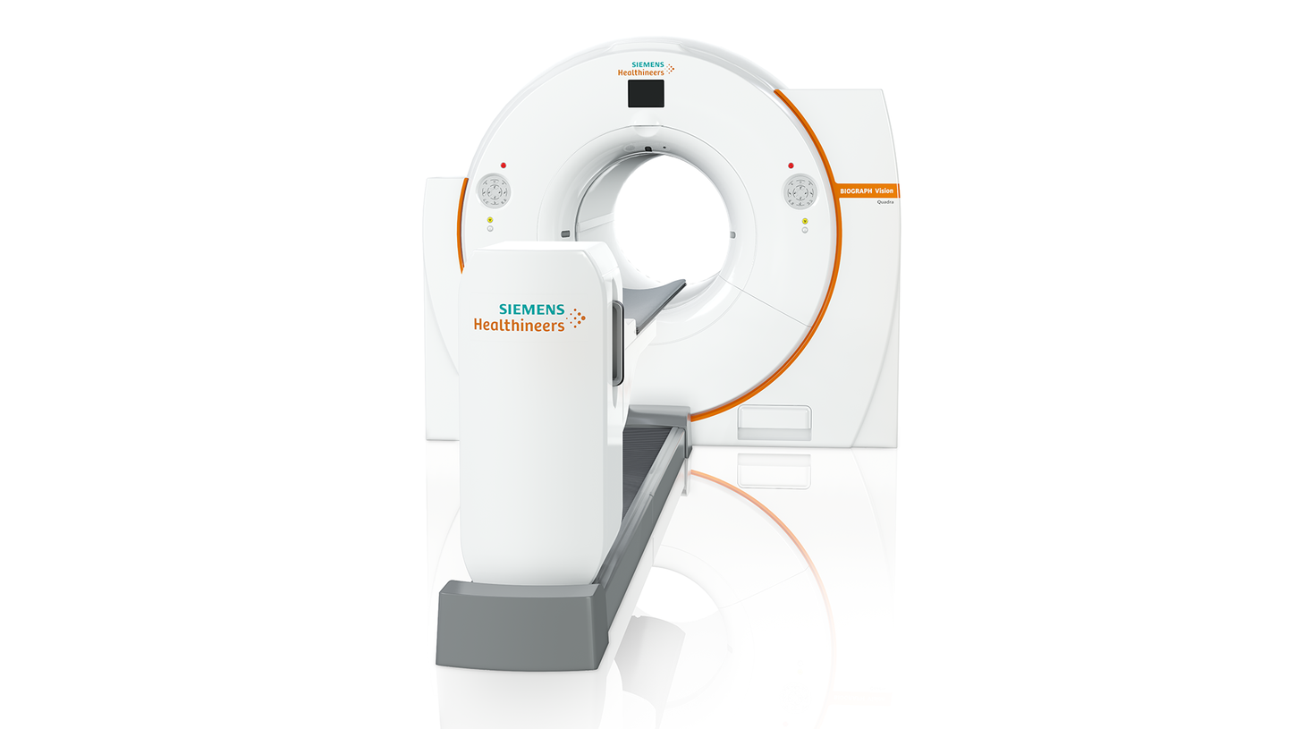
Biograph Vision Quadra6: Simultaneous whole-body imaging from head to thigh
The Biograph Vision Quadra PET/CT system expands the axial PET field of view to 106 cm, setting new standards in precision medicine. The system's larger axial field of view allows the nuclear physician to examine the patient over time during radiopharmaceutical imaging and also benefits from better anatomical coverage than a conventional PET/CT system. This type of faster scanning also results in lower radiation exposure for the patient.