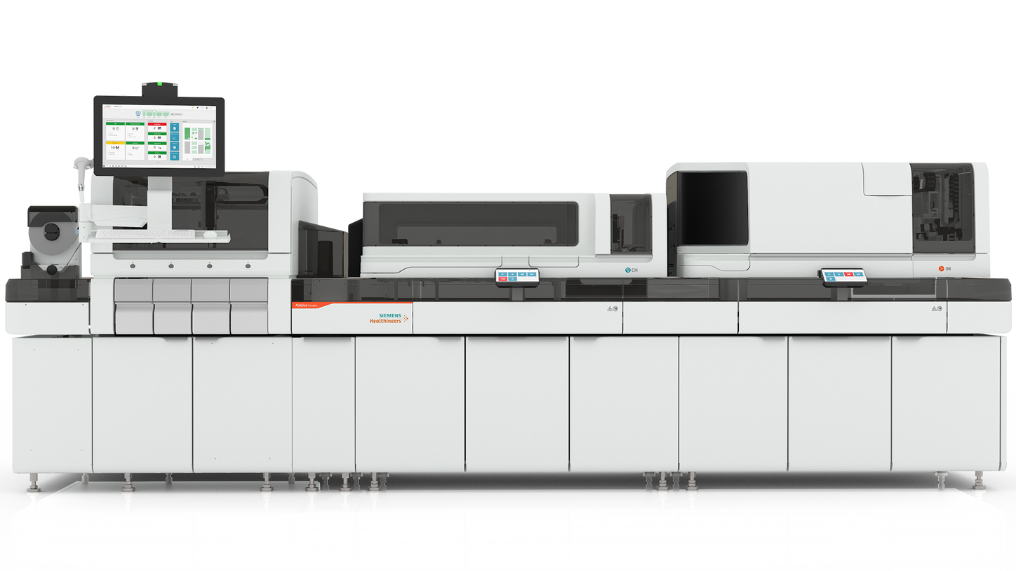
Current RUO Lineup
Advancement in neurology is not discovered by chance; it will be engineered through science. Siemens Healthineers is advancing neurodegenerative research by unlocking deep biological insight at the early stages of discovery. Our research assays are designed to help scientists better understand neuronal injury, disease progression, and the biological mechanisms that shape neurological health.
pTau2171
Tau is a microtubule associated protein found mainly in neurons, stabilizing microtubules and supporting neuronal structure. It is known to exist in multiple isoforms predominantly present within the central nervous system (CNS) and in the peripheral nervous system (PNS).
In neurodegenerative conditions, the tau protein may undergo changes, including hyperphosphorylation, a process associated with pathological changes in Alzheimer's disease.2 Our RUO pTau217 assay detects a low-molecular-weight hyperphosphorylated tau isoform thought to originate in the CNS that minimizes the peripheral signal supporting exploratory neurology research on Alzheimer's disease.3
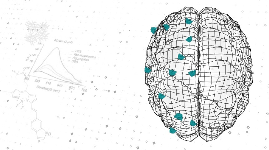
BDTau1
Brain-derived tau (BD-tau) is a form of the tau protein thought to originate specifically from neurons within the central nervous system. Tau normally stabilizes microtubules in axons, but when neurons are injured or degenerating, brain-specific tau fragments are released into circulation. BD-tau isolates these potentially CNS-derived tau species,3 which can be distinguished from peripheral tau isoforms. Research shows that this is similar to measuring total tau, but BD-tau levels may better reflect changes within the CNS due to neurodegenerative processes, such as those observed in dementia or due to acute injuries including stroke.4,5
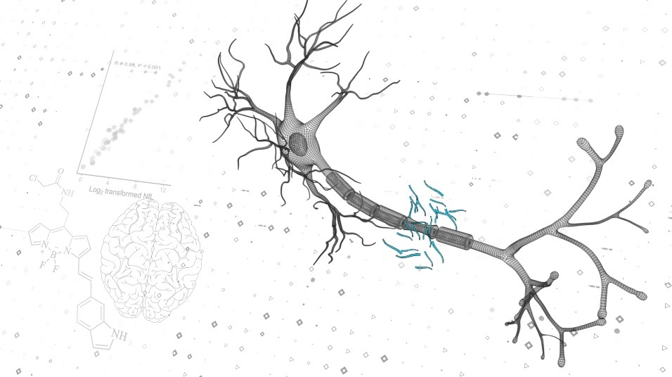
NfL6
Neurofilament light chain (NfL) belongs to a group of cytoskeletal elements called intermediate filaments specific to neurons. As intracellular structural components, these neurofilaments are found at lower levels in the blood of healthy individuals but can increase following neuroaxonal damage.7 Elevated NfL levels can occur as a result of multiple causes of nerve injury, including the neuroinflammatory axonal damage associated with multiple sclerosis-mediated myelin sheath destruction. These large-caliber, myelinated axons—commonly injured in multiple sclerosis—are a significant source of circulating NfL.8
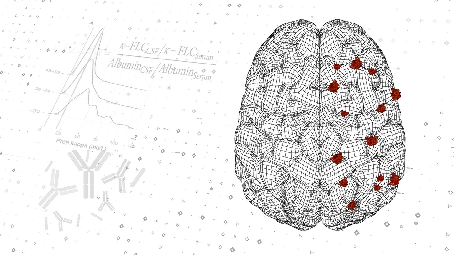
KFLC
Kappa free light chains (KFLC) are low‑molecular‑mass immunoglobulin components (~22–25 kDa) produced in slight excess during normal immunoglobulin synthesis and released as free molecules. KFLC are present in several biological fluids, including serum and cerebrospinal fluid (CSF). In published research, CSF KFLC concentrations have been observed to vary in cohorts studied in the context of central nervous system inflammation. KFLC measurements in CSF have been reported in neurological research studies, this has included exploratory research assessing how CSF KFLC measurements relate to oligoclonal band patterns generated by isoelectric focusing.
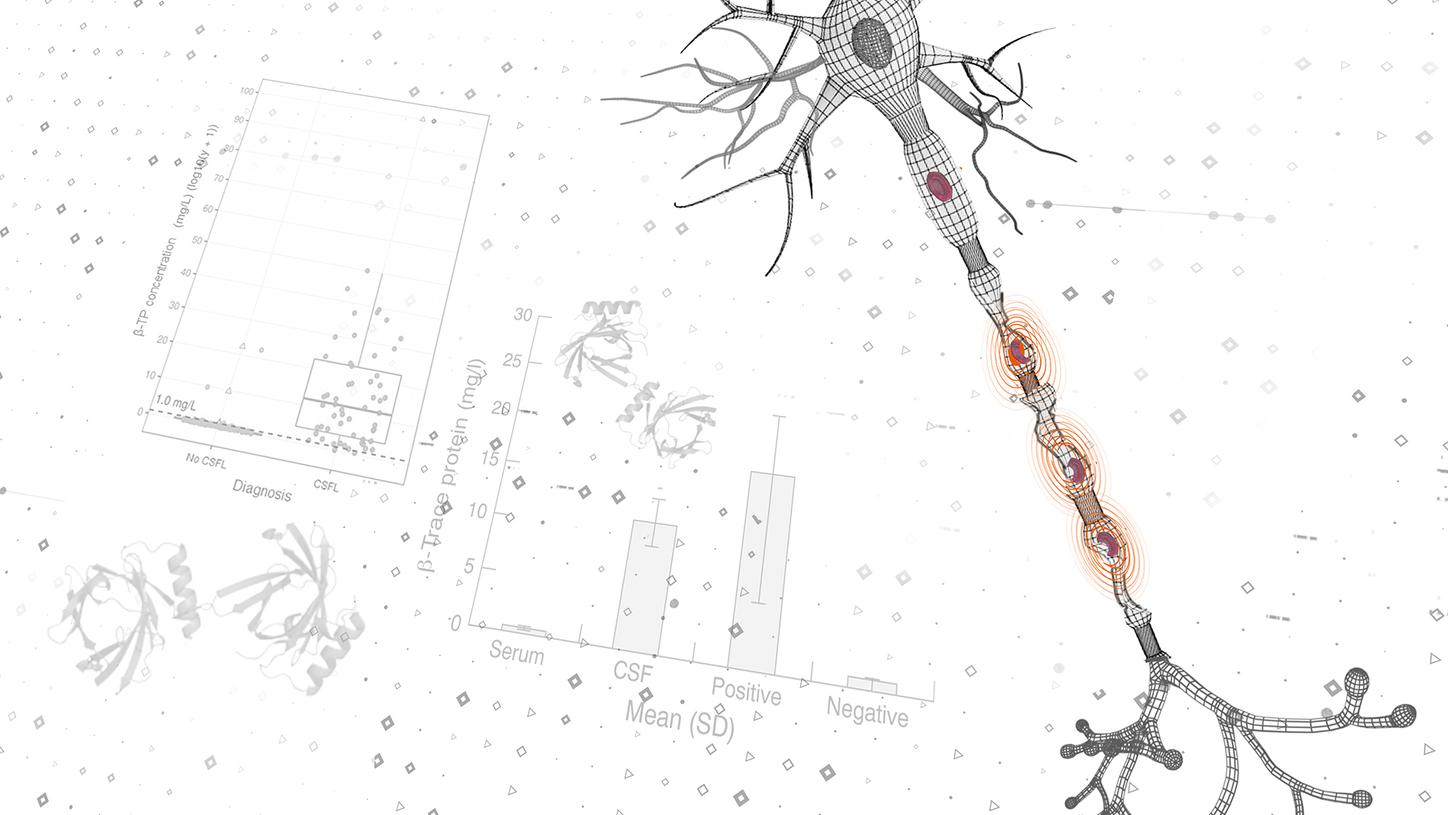
BTP
Beta trace protein (BTP), also known as prostaglandin D₂ synthase, is a low‑molecular‑ mass glycoprotein with an apparent molecular weight of approximately 23–29 kDa and is a member of the lipocalin protein family. BTP is predominantly synthesized within the central nervous system by glial cells and the choroid plexus and constitutes one of the principal protein components of cerebrospinal fluid (CSF). BTP expression has been described in most tissues, with the exception of the ovaries, and the protein can be detected in multiple biological fluids.
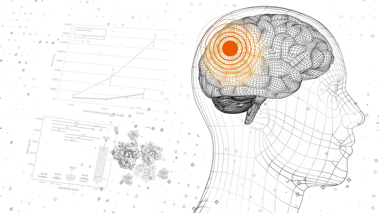
SAA
Serum amyloid A (SAA) is an acute‑phase protein primarily synthesized by the liver in response to pro‑inflammatory cytokines released by activated monocytes. SAA can also be produced by macrophages, endothelial cells, smooth muscle cells, adipocytes, and other cell types at sites of inflammation. Plasma SAA concentrations can increase markedly following an inflammatory stimulus, with initial elevations observed within hours and peak levels reached within a few days. SAA has been extensively described in inflammation research.
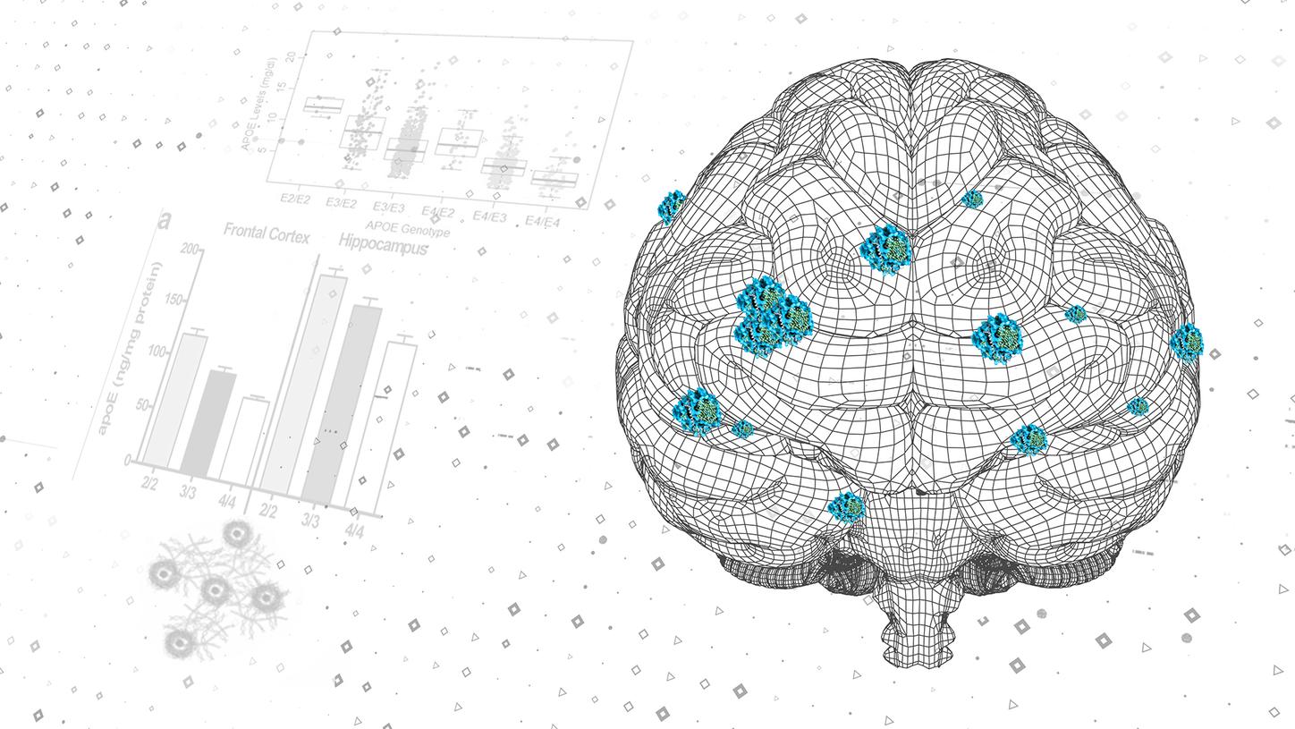
Apo E
Apolipoprotein E (ApoE) is a polymorphic protein with a molecular weight of approximately 34kDa (299 amino acids) that is synthesized primarily in the liver. Apo E exists in three major isoforms—Apo E2, Apo E3, and Apo E4—with plasma concentrations described to vary according to the dominant isoform present. ApoE interacts with low‑density lipoprotein receptors and other proteins and plays a role in the transport and metabolism of cholesterol and lipids, constituting a significant component of very‑low‑density lipoproteins. Variations in Apo E levels and isoform distribution have been widely described in lipid metabolism and neurovascular research contexts.