Dual Source CT in cardiac imaging
Explore untapped potential
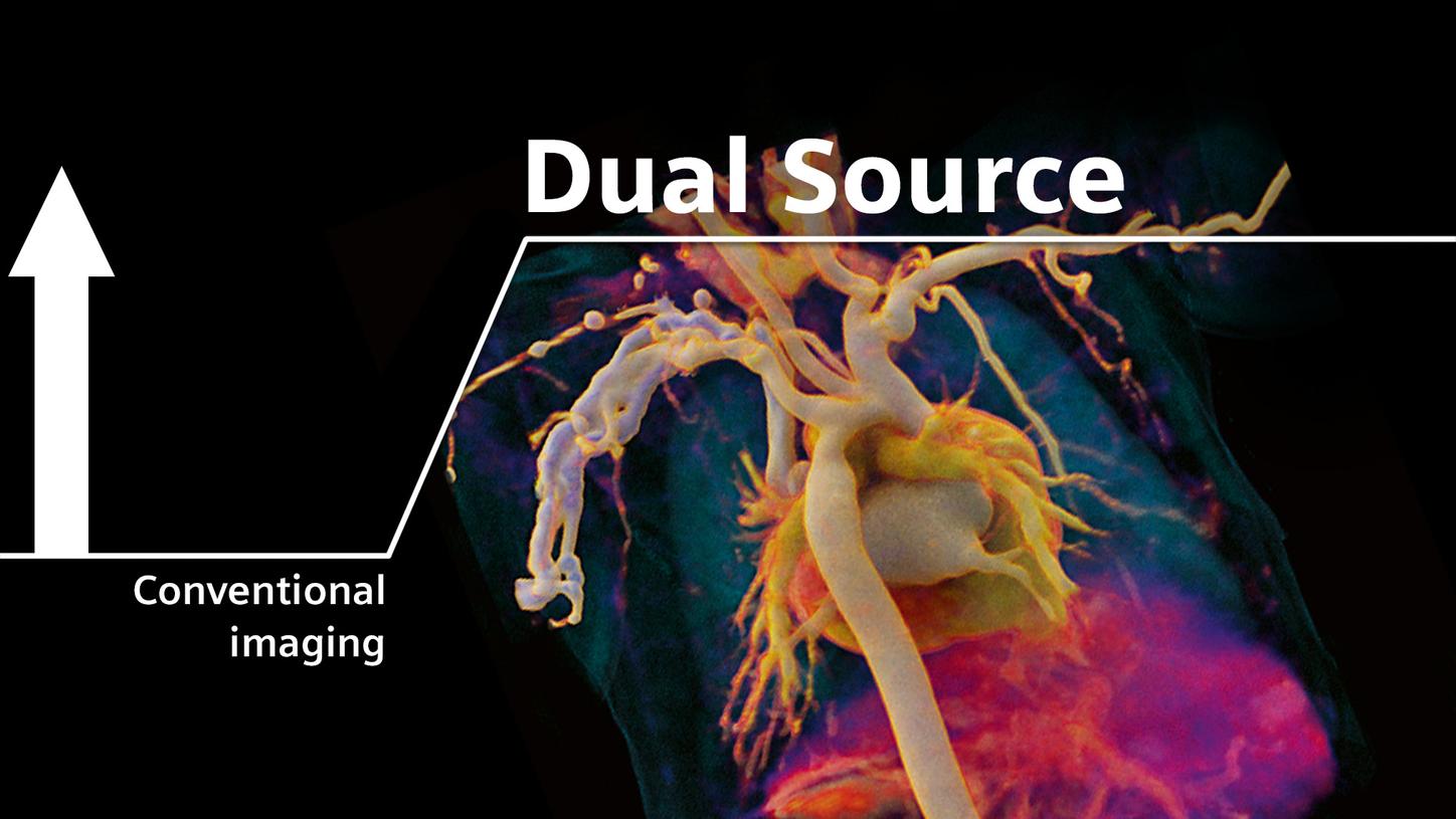
Cardiac CT was the starting point of Dual Source CT – and users have employed Dual Source technology to push boundaries ever since. With a world of applications to explore, how can we help you tap into the full potential of Dual Source CT?
Did this information help you?
Suh YJ, et al. (2019): Tricuspid annular diameter and right ventricular volume on preoperative cardiac CT can predict postoperative right ventricular dysfunction in patients who undergo tricuspid valve surgery . Int J Cardiol. 2019;288:44-50.
Hashimoto G, Fukui M, Sorajja P, Cavalcante JL. Essential roles for CT and MRI in timing of therapy in tricuspid regurgitation. Prog Cardiovasc Dis. 2019;62(6):459–62.
Abadia AF, van Assen M, Martin SS, Vingiani V, Griffith LP, Giovagnoli DA, et al. Myocardial extracellular volume fraction to differentiate healthy from cardiomyopathic myocardium using dual-source dual-energy CT. J Cardiovasc Comput Tomogr. 2019;(September).
See, e.g., Pontone G, et al. (2019): Determinants of Rejection Rate for Coronary CT Angiography Fractional Flow Reserve Analysis
Li W, Yu F, Zhu W, Zhang W, Jiang T. Detection of left atrial appendage thrombi by third-generation dual-source dual-energy CT: Iodine concentration versus conventional enhancement measurements. Int J Cardiol [Internet]. 2019
Han BK, et al. (2015): Computed Tomography Imaging in Patients with Congenital Heart Disease Part I: Rationale and Utility. An Expert Consensus Document of the Society of Cardiovascular Computed Tomography (SCCT).
Barrera CA, et al. (2018): Depiction of the native coronary arteries during ECG-triggered High-Pitch Dual-Source Coronary Computed Tomography Angiography in children: Determinants of image quality