
syngo DynaPBV BodyEvaluate perfusion for personalized therapy
syngo DynaPBV Body is a software application which complements 3D anatomical imaging with volumetric physiological information directly in the interventional lab.
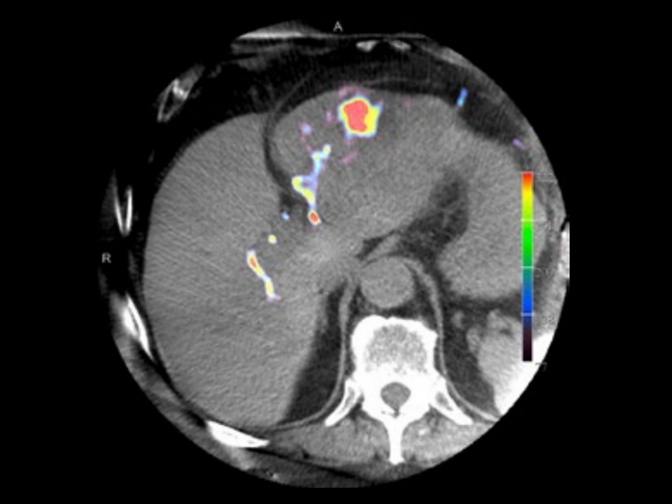
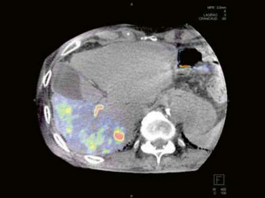
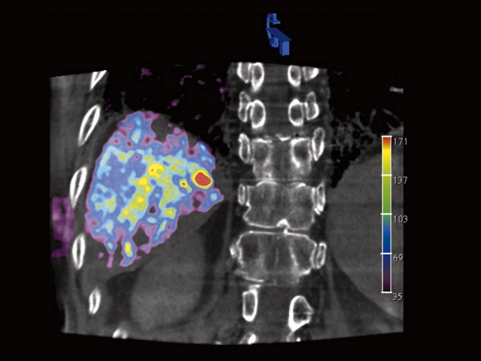
The software indicates the distribution of blood in lesions and surrounding tissue by means of color-coded cross-sectional blood volume maps. Based on this blood volume information, physicians can evaluate changes in perfusion caused by treatment or biological processes. It can be used to monitor response to treatment following repeated TACE and has the potential to identify potential non-responders directly intra-procedurally.
Features & Benefits

Features
- Allows for blood volume measurements
- Makes physiological information available directly in the angio suite
- Provides high-resolution contrast-enhanced syngo DynaCT images for delineation of tumor-feeding vessels as well

Benefits
- Determine the specific characteristics of each individual lesion
- Identify potential non-responders directly intra-procedurally
- Facilitate control of the optimal end point of your intervention
Clinical Use
Hear what our customers are saying1
Interview with Prof. Gerd Groezinger
Precision in embolization- boosted by syngo DynaPBV.
As a strong believer of the regular use of syngo DynaCT, Prof. Gerd Groezinger performs syngo DynaPBV scans routinely during TACE procedures. Learn how syngo DynaPBV scans serve two crucial purposes:
- identifying tumor feeders for effective planning
- determining the need for immediate on-table embolization or rescheduling based on residual tumor perfusion.
"syngo DynaPBV guidance led to a reduced number of TACE-interventions without negative impact on response or survival"2

Prof. Dr. Gerd Grözinger
Interventional Radiology University Hospital Tuebingen
syngo DynaPBV Body
Click through the clinical cases

Thomas Vogl, MD, Diagnostic and Interventional Radiology, University of Frankfurt, Germany
High-resolution contrast-enhanced syngo DynaCT images provided by syngo DynaPBV Body.
Courtesy: Prof. Gerd Groezinger, Interventional Radiology, University Hospital Tuebingen, Germany
syngo DynaPBV Body blood volume map only

Courtesy: Prof. Vogl, Dept. of Diagnostic and Interventional Radiology, University of Frankfurt, Germany
Blood volume map overlaid on native syngo DynaCT images provided by syngo DynaPBV Body.

Courtesy: Prof. Vogl, Dept. of Diagnostic and Interventional Radiology, University of Frankfurt, Germany
Blood volume map overlaid on native syngo DynaCT images provided by syngo DynaPBV Body.

Courtesy: Prof. Vogl, Dept. of Diagnostic and Interventional Radiology, University of Frankfurt, Germany
Blood volume map overlaid on native syngo DynaCT images.

Courtesy: Thomas Vogl, MD, Diagnostic and Interventional Radiology, University of Frankfurt, Germany
Blood volume map overlaid on native syngo DynaCT images provided by syngo DynaPBV Body.

Thomas Vogl, MD, Diagnostic and Interventional Radiology, University of Frankfurt, Germany
High-resolution contrast-enhanced syngo DynaCT images provided by syngo DynaPBV Body.


Papers & Studies
Case Studies & Study Protocols

Transarterial chemoembolization of a hepatocellular carcinoma in the liver
Courtesy of Prof. G. Grözinger, MD, A. Estler, MD, Interventional Radiology Department, University Hospital Tübingen, Germany

Comprehensive Tumor Assessment as the Foundation of Optimal Therapy Planning
Courtesy: Masao Hamuro, MD, Yoshinori Takao, and Takao Ichida, Osaka City University Hospital, Japan

Transarterial chemoembolization of HCC using syngo DynaPBV Body
Courtesy of Jeff McCann, MD, Ronan Ryan, MD, Department of Interventional Radiology, St. Vincent University Hospital, Dublin, Ireland

Transarterial chemoembolization of HCC
Courtesy: Masao Hamuro, MD, Yoshinori Takao, and Takao Ichida, Osaka City University Hospital, Japan
Scientific Talks and Publications
Link | Title | Authors |
Usefulness of Cone-Beam CT-Based Liver Perfusion Mapping for Evaluating the Response of Hepatocellular Carcinoma to Conventional Transarterial Chemoembolization. | Choi SY, Kim KA, Choi W, Kwon Y, Cho SB. | |
Intraprocedural cone-beam CT with parenchymal blood volume assessment for transarterial chemoembolization guidance: Impact on the effectiveness of the individual TACE sessions compared to DSA guidance alone. | Peisen F, Maurer M, Grosse U, Nikolaou K, Syha R, Artzner C, Bitzer M, Horger M, Grözinger G. | |
Transarterial Chemoembolization of Hepatocellular Carcinoma Using Radiopaque Drug-Eluting Embolics: How to Pursue Periprocedural Cross-Sectional Imaging? | Ruff C, Grözinger G, Syha R, Elser S, Partovi S, Bitzer M, Horger M, Nikolaou K, Grosse U. | |
Correlation of C-arm CT acquired parenchymal blood volume (PBV) with 99mTc-macroaggregated albumin (MAA) SPECT/CT for radioembolization work-up. | Weissinger M, Vogel J, Kupferschläger J, Dittmann H, Castaneda Vega SG, Grosse U, Artzner C, Nikolaou K, la Fougere C, Grözinger G. | |
Intraprocedural Parenchymal Blood Volume Is a Predictor of Treatment Response for Chemoembolization in Hepatocellular Carcinoma: Results of a Prospective Study. | de Korompay N, Alshammari M, Klass D, Chou FY, Chung J, Ho S, Liu DM. | |
Image quality of arterial phase and parenchymal blood volume (PBV) maps derived from C-arm computed tomography in the evaluation of transarterial chemoembolization. | Zitzelsberger T, Syha R, Grözinger G, Partovi S, Nikolaou K, Grosse U. | |
Intraprocedural 3D perfusion measurement during chemoembolisation with doxorubicin-eluting beads in liver metastases of malignant melanoma. | Pereira PL, Krüger K, Hohenstein E, Welke F, Sommer C, Meier F, Eigentler T, Garbe C. | |
Quantitative assessmant of gastric perfusion following bariatric arterial embolization in porcine model. | Genyk P, Ehtiati T, Paudel K, Krimins R, Akinwade O, Arepally A, Kraitchman D, Weiss C. | |
Assessment of the parenchymal blood volume by C-arm computed tomography for radioembolization dosimetry. | Grözinger G, Kupferschläger J, Dittmann H, Maurer M, Grosse U, la Fougère C, Nikolaou K, Syha R, Ketelsen D. | |
Quantitative liver tumor blood volume measurements by a C-arm CT post-processing software before and after hepatic arterial embolization therapy: comparison with MDCT perfusion. Diagn Interv Radiol. 2015 Jan-Feb;21(1):71-7.doi: 10.5152/dir.2014.13290. PMID: 25538037; PMCID: PMC4463359. | Peynircioğlu B, Hızal M, Çil B, Deuerling-Zheng Y, Von Roden M, Hazırolan T, Akata D, Özmen M, Balkancı F. |
Avez-vous jugé cette information utile?
Merci beaucoup
1) The statements by Siemens Healthineers' customers described herein are based on results that were achieved in the customer's unique setting. Since there is no "typical" hospital and many variables exist (e.g., hospital size, case mix, level of IT adoption) there can be no guarantee that other customers will achieve the same results.
2) Source: Peisen F, Maurer M, Grosse U, et al. Eur J Radiol. 2021;140:109768. The analysis of the influence of intraprocedural DynaPBV was based on matched pair analysis (DynaPBV n = 28 vs. DSA n = 28) in a retrospective single center study.