keyvisual teamplay X-ray Dashboard

teamplay X-ray DashboardIncrease quality of imaging through transparency

How do you figure out why X-ray images are being rejected at your imaging department and identify areas which need improvement?

How do you make sure radiation dose is applied correctly and your patients are not exposed to levels of radiation dose that are higher than necessary whilst still ensuring high quality of X-ray images?

How do you stay compliant with the ever-increasing requirements for exam patient documentation?

  • Provides transparency on imaging data, including full rejected analysis, helping to boost efficiency
  • Identifies variances in imaging, allowing you to take measures to reduce radiation dose
  • Tracks and documents rejection rate and imaging variances, letting you comply with exam documentation guidelines
Customer voices

Vesalius

Our solution for reject analysis

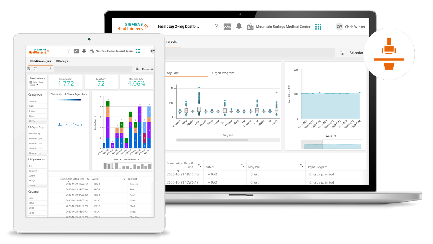
teamplay X-ray Dashboard2 provides you with a full analysis of rejected images, identification of the root cause of the rejection and an overview of variances in imaging based on exposure index (EXI) values.

It also helps you comply with strict guidelines for exam documentation. If you can identify inefficiencies, improvements can be made. Achieve more transparency and boost your efficiency.

teamplay X-ray Dashboard - Reject analysis

teamplay X-ray Dashboard – Reject Analysis

Deep dive into reject analysis with the dedicated dashboard. Monitor your overall rejection rate at a glance and analyze possible causes of rejected images for reducing image retakes. Use additional filters like body regions, organ programs, operator names or specific systems to identify appropriate measures to reduce the rejection rate and the radiation dose across your X-ray department. Reducing the retake rate can also result in higher patient throughput.  

teamplay X-ray Dashboard  EXI Analysis

teamplay X-ray Dashboard – EXI Analysis

If you notice significant deviations from target levels in the EXI value, which could indicate systematic over- or underexposures, you can drill down to filter on body region, organ program, operator name or a specific system. These insights enable you to accordingly derive corrective actions like modifying the organ program or educating your staff. This ensures that the patient is not exposed to excessive levels of radiation and with this, helps in improving the quality of X-ray examinations.

1
2
3