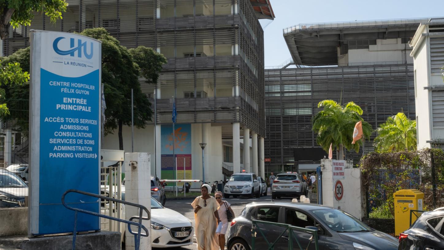
This Indian Ocean island is recognized for its natural beauty and vibrant culture, offering a range of outdoor activities and culinary experiences. However, beyond its appeal to visitors, the island is making significant strides in healthcare. The installation of a total- body PET/CT scanner at Centre Hospital Universitaire is elevating the standard of medical care for residents and neighboring islands, supporting both clinical and research needs.
Photography by Romain Philippon | Data courtesy of Centre Hospital Universitaire, La Réunion.
Access to PET/CT diagnostics on the French island of Reunion Island, also referred to as La Réunion, situated between Madagascar and Mauritius in the Indian Ocean, has seen a dramatic transformation. With a surge in the number of patients able to benefit from advanced scans and a growing range of examinations to offer, the island’s medical landscape is buzzing with possibility.
Cédric Revel, MD, is head of the department of nuclear medicine at Centre Hospital Universitaire, a tertiary care provider that serves all the island’s 900,000 inhabitants. The hospital also takes on patients from Mauritius, Mayotte, and other islands in the vicinity.
“Until recently, we could not offer PET/CT to more than 3,000 patients per year. Waiting time was typically about 6 weeks,” says Revel. Something had to be done. The existing scanner, Biograph mCT, was simply not enough anymore. After discussion, the decision was taken to go big: in 2023, Biograph Vision Quadra PET/CT was put into operation.1
For Revel, this marked the beginning of a new molecular imaging era on La Réunion. “We were able to more than double the number of patients scanned to about 8,000 per year. Waiting time is down to two or three days, which means that access to PET/CT on La Réunion is better than in many parts of France.” About 97% of the PET/CT scans are done with Biograph Vision Quadra now. Biograph mCT continues to support the department as a backup scanner.
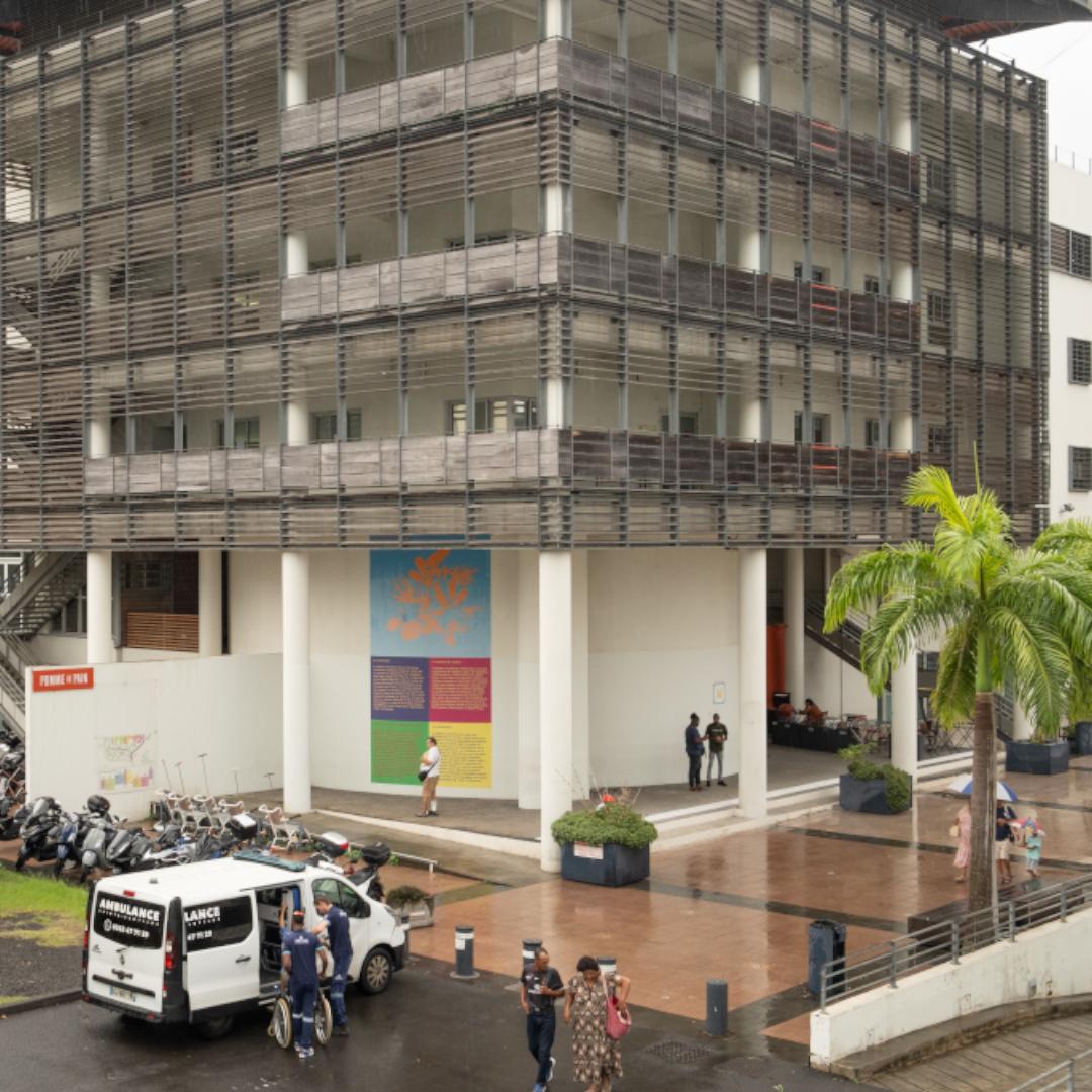
Centre Hospital Universitaire, La Réunion.
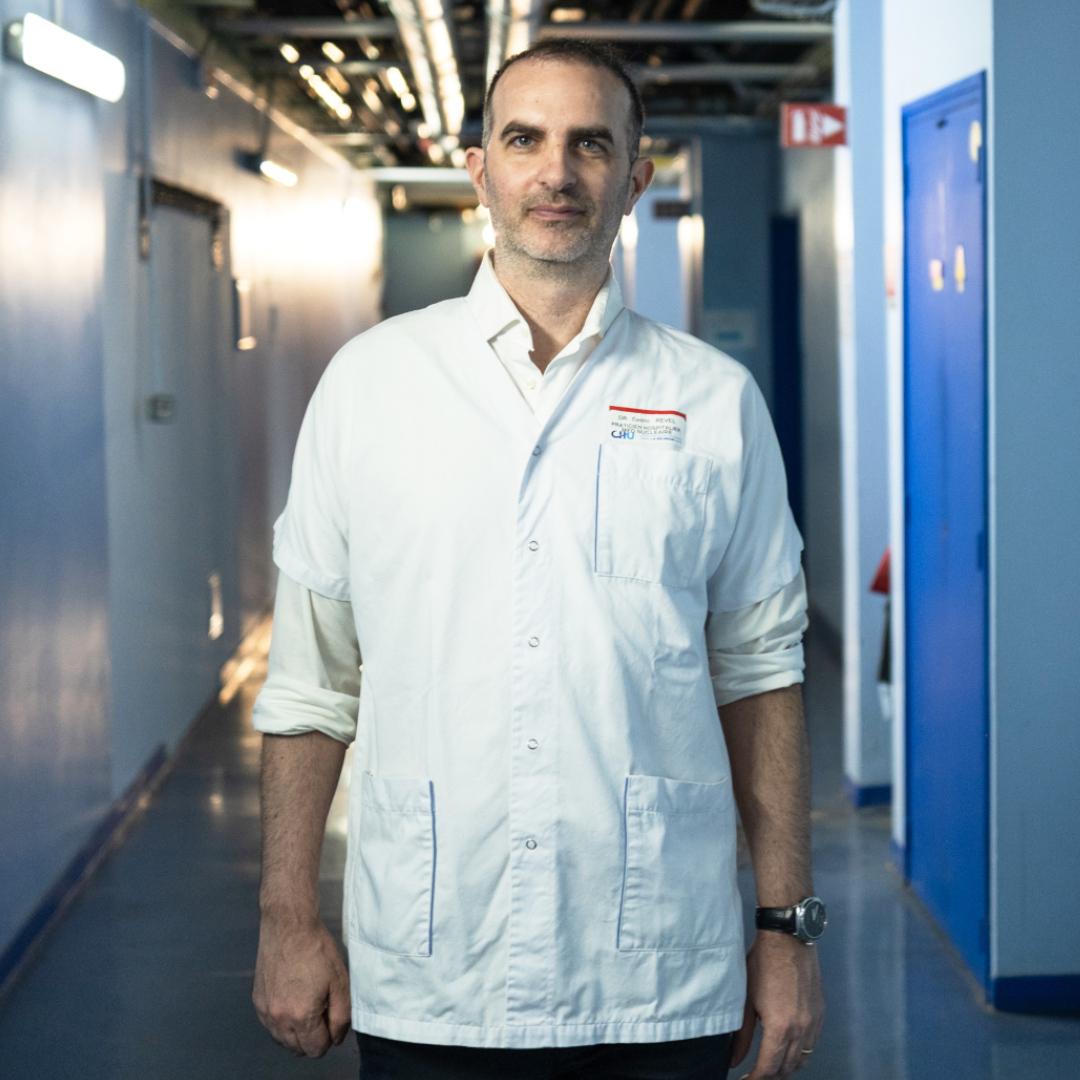
Cédric Revel, MD, Centre Hospital Universitaire, La Réunion.
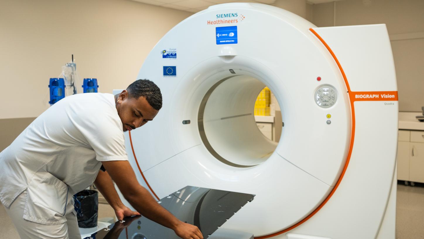
Technician adjusts Biograph Vision Quadra PET/CT's radiation therapy pallet.
Collaboration with France
Being able to perform 8,000 examination a year is one thing. Being able to interpret all of these scans is a second challenge. Shortage of physicians is an issue, which is why the department is cooperating with IMADIS, one of the largest teleradiology providers in France. PET/CT and SPECT images can be transferred securely in a digital imaging and communications in medicine (DICOM) format to mainland France, and the interpretation comes back within a few days. Revel is more than happy to have this option available. “The volume varies depending on staff situation. Sometimes it is 20 patients per week, while sometimes it is 20 per day.”
PET/CT also requires technologists, of course. This is less of an issue, says Mourad Baiba, who is working as a technologist both in nuclear medicine and in radiology. “We have a school for technologists on the island. Our department is highly ranked among the students. Many want to work here.” With the introduction of Biograph Vision Quadra PET/CT, workflows have changed, according to Baiba. “We examine patients really quickly nowadays, so we need more technologists than before.” And they must be highly qualified, since seemingly short delays of one to two minutes per patient quickly add up when more than 50 patients are scanned using the total-body PET/CT and more than 55 PET examinations are performed each day.
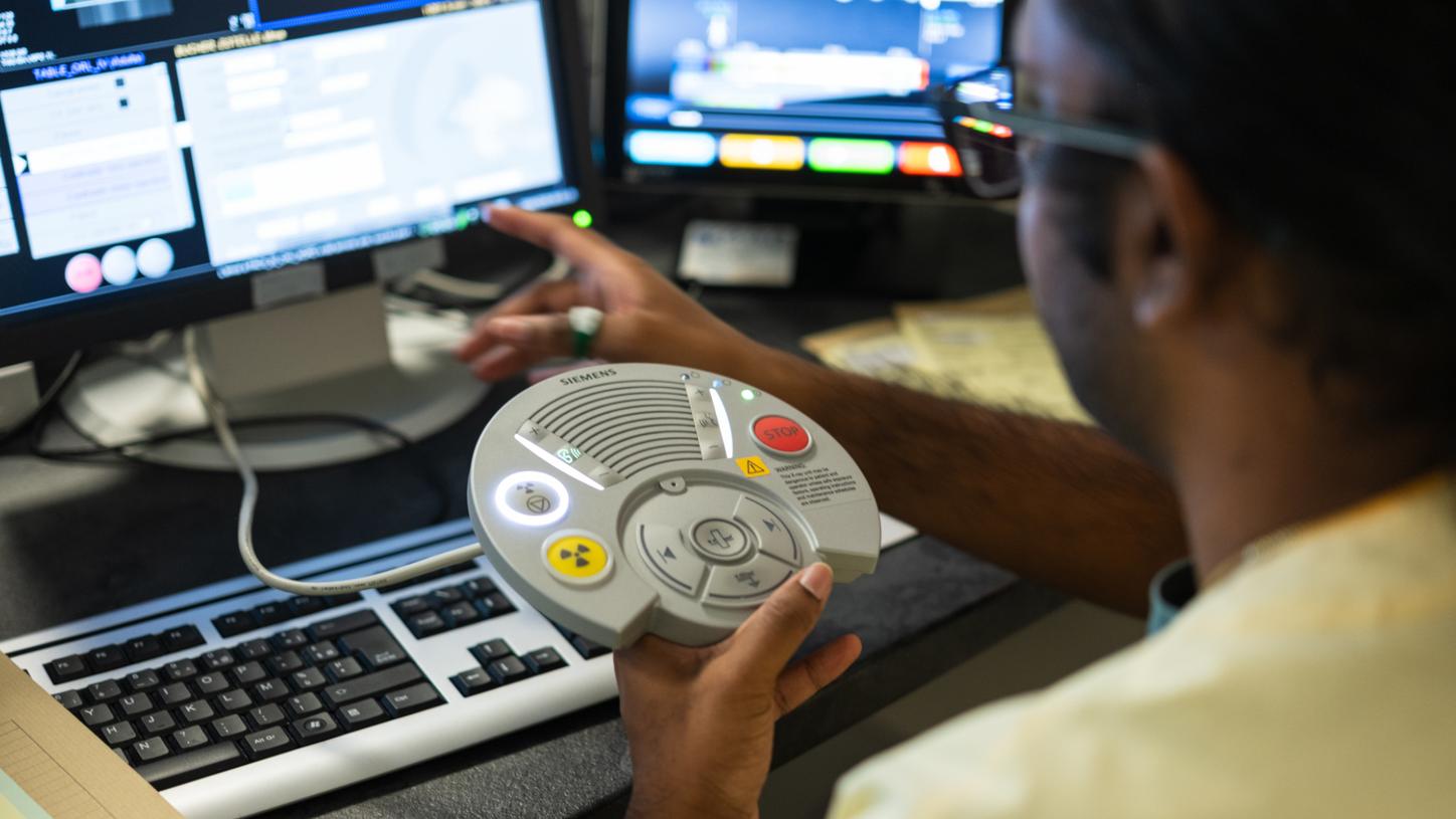
This device is used to communicate with the patient, move the patient bed, and start the radiation of the CT.
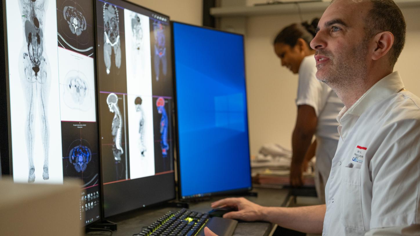
Revel reviewing images in the control room.
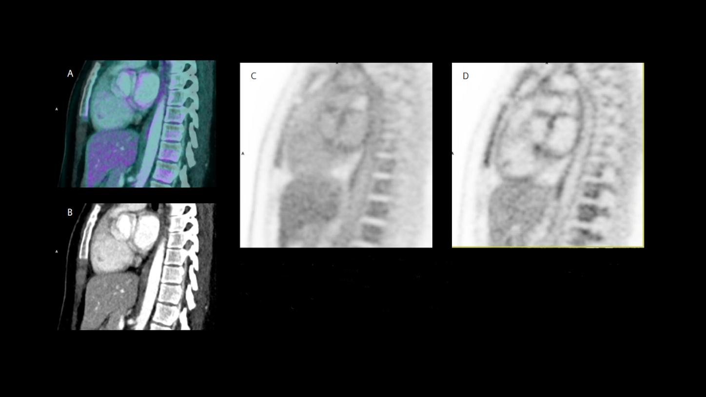
Example of an endometriosis research protocol of a patient in their mid 30s for assessment of the cardiac walls. Administered dose of 3.5 MBq/kg of 18F-FDG.2, 3 Images were acquired with a scan time of 10 minutes.
"PSMA scans with Biograph Vision Quadra are not only easy to do, but they are also very cost-effective."
Unexpected patient benefit
For Revel, the two most remarkable things about this total-body PET/CT from a medical point of view are its high sensitivity and resolution. “We didn’t expect that, but the resolution of the PET part is so good that, in many cases, we do not need a contrast agent for a CT scan anymore like we did with our previous scanner. The combination of higher resolution and very low image noise on this scanner has made scans much more sensitive, which means that we rely less on iodine-enhanced CT than we used to. We still maintain contrast-enhanced CT for the abdominopelvic region and for head-and-neck imaging when vascular relationships need to be assessed, but contrast-enhanced computed tomography (CECT) now adds less value than it did previously. As a result, we have reduced our use of CECT by more than 60%, whereas it was previously almost systematic.”
To achieve the best possible sensitivity in oncological PET/CT, the department uses a hydration protocol that requires patients to drink two glasses of water before fludeoxyglucose injection F-18 (18F-FDG) injections. Revel observed natural tissue radioactivity was reduced significantly using this protocol, especially in the liver, which made it easier for him to see lesions.
The high sensitivity is also what has allowed Revel and his team to expand PSMA imaging in prostate cancer. “PSMA scans with Biograph Vision Quadra are not only easy to do, but they are also very cost-effective. With two runs, we can scan 16 PSMA-patients per day.” Centre Hospital Universitaire’s standard dose is between 0.5 and 0.6 MBq/kg per injection, way less than required with conventional scanners.2, 3
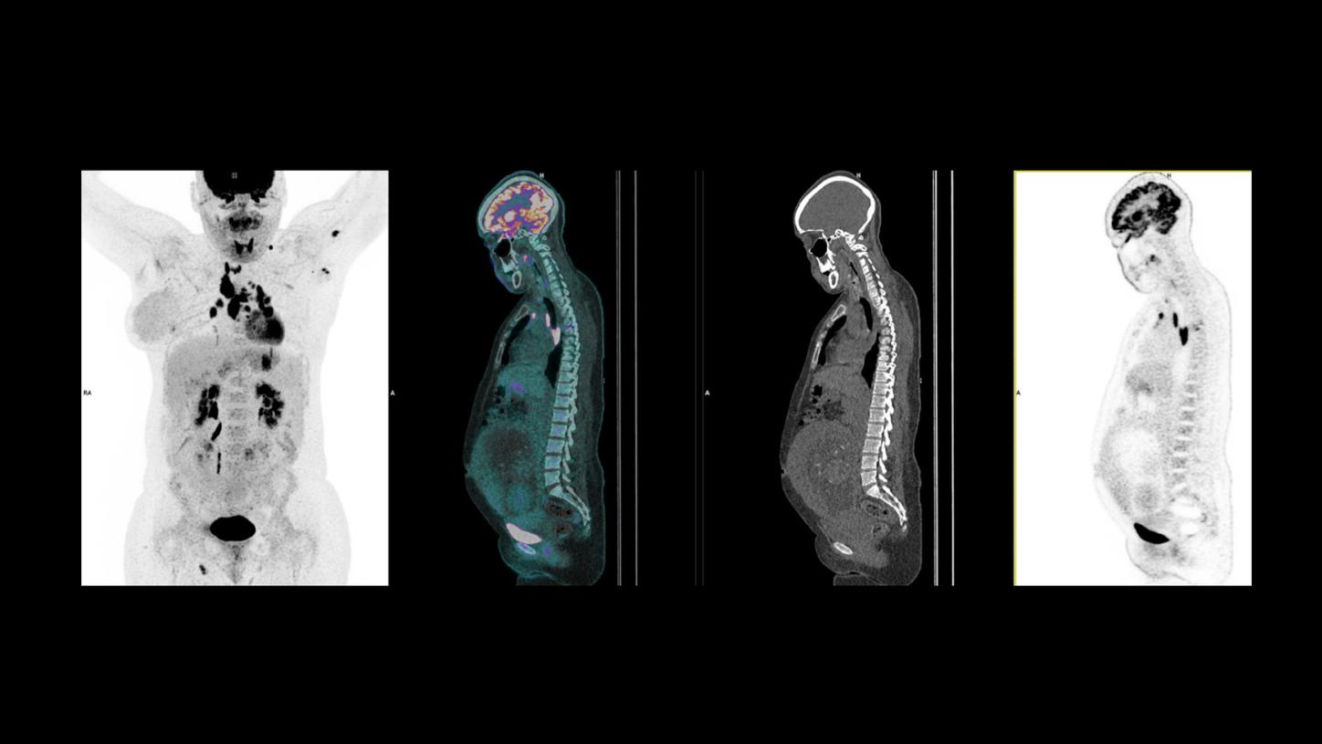
Pregnant (20 weeks); Breast Cancer | CT: Tin filter; DLP: 45 mGy | 84 kg; 1.65 m; BMI: 31 | Dose: 0.5 | MBq/kg 18F-FDG2, 3: 44 MBq, Acquisition: 10 min
Island’s cyclotron helps broaden clinical spectrum
Revel is now planning to expand the spectrum of examinations that his department can offer. The island’s cyclotron, established in 2008, has been used mainly for 18F-FDG. The plan for 2026 is to add 18F-Fluorocholine (18F-FCH) and 18F-Fluorodopa (18F-DOPA). 18F-FCH is used for parathyroid imaging, which, according to Revel, is a large improvement to the 4D-CT scans currently being used in their SPECT/CT studies. 18F-DOPA is for PET/CT imaging in Parkinson’s disease, and it will also be helpful for cerebral metastases.
A third tracer that will be available on La Réunion from 2026 onwards is Rubidium-82, a potassium-mimetic for myocardial perfusion imaging. Currently myocardial perfusion imaging is done with SPECT/CT, approximately 4,000 examinations per year. Revel is planning to move some of these over to the PET/CT—given that PET/CT based myocardial perfusion imaging has high diagnostic accuracy and allows quantification of myocardial blood flow.
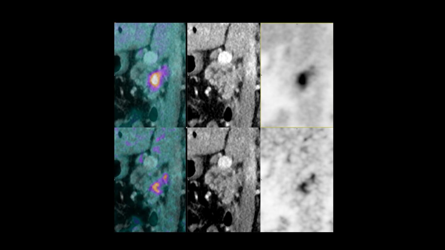
Pancreatic cancer, staging workup; FlowMotion AI (continuous bed motion).
Total scan duration 6 minutes, with a dedicated zone over the pancreases for high-resolution imaging of 2 minutes. Bottom row of images emphasis the high-resolution capability and use of OncoFreeze AI motion management over the pancreas. This allowed for the visualization of a lymph node located behind the pancreatic tumor. This lymph node was only clearly seen on the very high-resolution image.
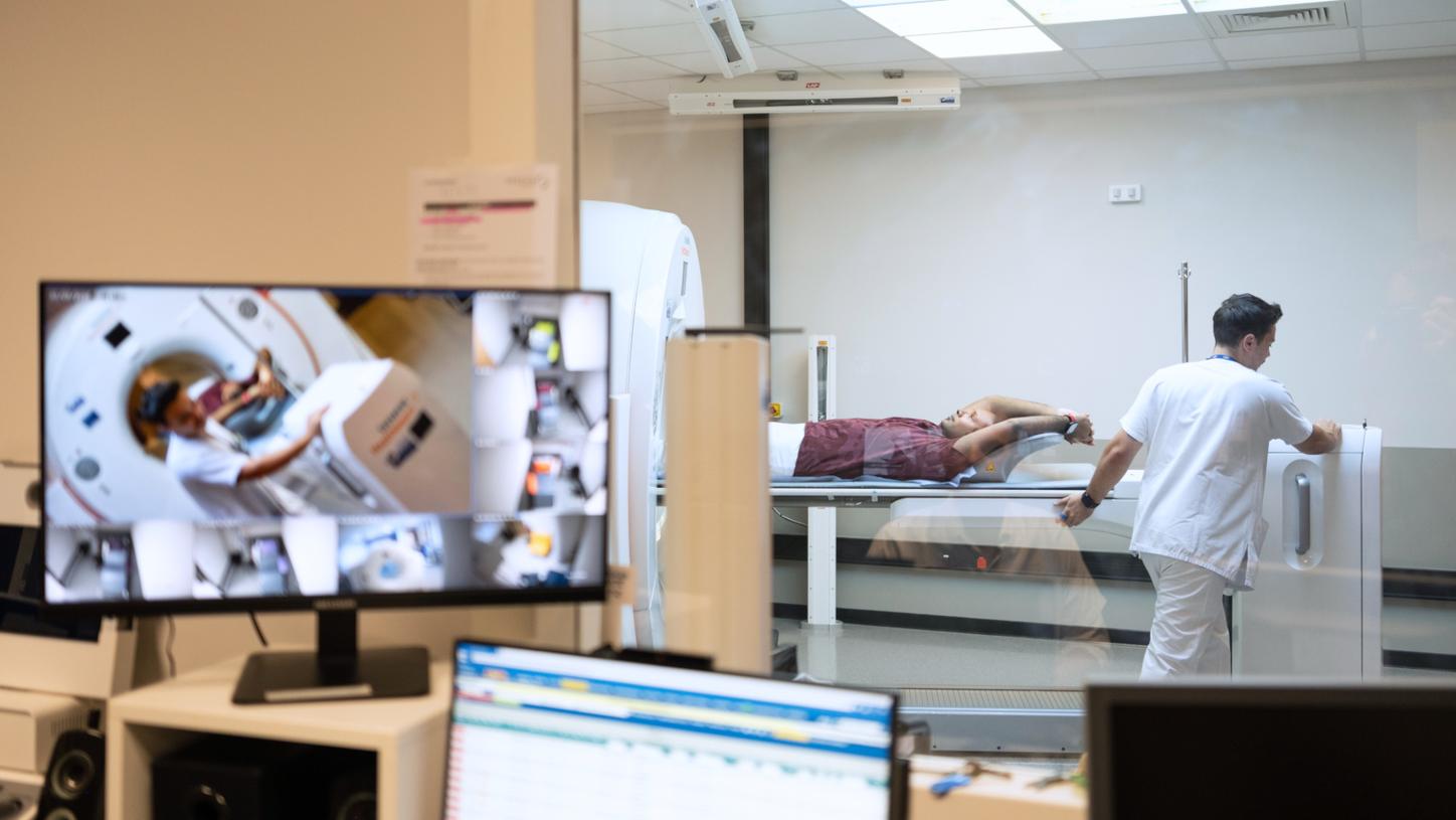
Control room view of the Biograph Vision Quadra PET/CT.
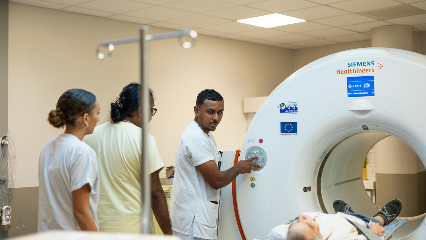
Nuclear medicine team members at Centre Hospital Universitaire.
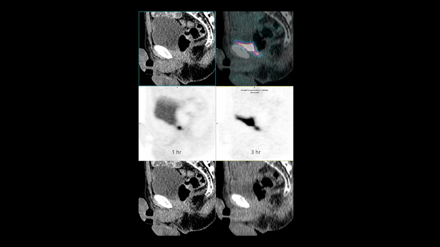
Research ambitions in field of screening
And then there is Fibroblast Activation Protein Inhibitor (FAPI). (18F)-FAPI is a relatively new tracer shown to have potential use in indications alternative to 18F-FDG for oncological imaging. It is extremely sensitive, and the low radiation doses needed make it highly suitable for oncological screening examinations. This is a field that Revel wants to explore further. “In recent years, PET/CT research has been very difficult for us because of the long waiting times. Thanks to this total-body PET/CT, we have capacity now, and we will use this extra time on research in the field of (18F)-FAPI screening.”
Revel articulates, “When it comes to screening, the standard reflex in the medical community has long been that PET/CT should not be considered in asymptomatic individuals because of radiation exposure. However, with the advent of modern scanners and low-dose tracers, we believe research should not overlook clinical studies exploring whether PET could help with multiple cancer early detection (MCED) and chronic diseases in asymptomatic patients. Our approach remains strictly research-based and phased, beginning with proof-of-concept studies; only if these are conclusive would a future cohort study be considered.” Possessing this receptive outlook, the island’s medical offerings are sure to expand and benefit the community.

Centre Hospital Universitaire
About the author
Philipp Grätzel is a medical doctor by training, who has been working as a healthcare and technology journalist in Berlin for many years.


