Abstract
Extensive coronary artery calcification (CAC) remains a major limitation in coronary CT angiography (CCTA), often resulting in non-diagnostic studies due to blooming artifacts and impaired lumen visualization1, particularly in patients with calcium scores exceeding 1,000. The implementation of Photon Counting Detector CT (PCD-CT (NAEOTOM Alpha) represents a significant advancement in overcoming these barriers2.
This case report describes a patient with an Agatston score of 11,0003, where the PCD-CT enabled clear delineation of the coronary lumen and confident assessment of stenosis without the need for invasive catheter angiography. By significantly reducing blooming artifacts and enhancing spatial resolution, PCD-CT is pushing the boundaries of non-invasive cardiac imaging, breaking through the limitations of conventional Energy-Integrating Detector (EID) CT4, and establishing a new benchmark for evaluating patients with extreme calcific burden. The future of cardiac care just got brighter!
Case Presentation
A 62-year-old male with poorly controlled type II and Hypercholesterolemia, an active lifestyle, no cardiac or chest symptoms, underwent an annual health checkup, and low-dose CT lung screening showed extensive coronary artery calcification. The patient was subsequently referred for CCTA to further evaluate coronary artery disease risk. Non-contrast calcium scoring revealed an Agatston score of 11,000, indicating an extremely high calcific burden.
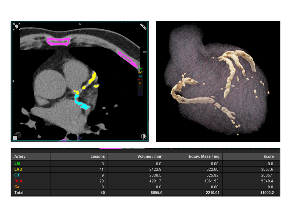
Fig. 1. Quantitative summary of calcium scoring with a cumulative Agaston score of 11,000, reflects a high burden of calcification
To overcome the diagnostic limitations typically associated with such extensive calcification, the patient was scanned using the Quantum Plus HD protocol on the NAEOTOM Alpha system. The ultra high-resolution PCD-CT acquisition with spectral data reconstruction6 enabled clear visualization of the coronary lumen and confident assessment of stenosis, despite the dense calcific burden. Invasive catheter angiography was avoided in this case.
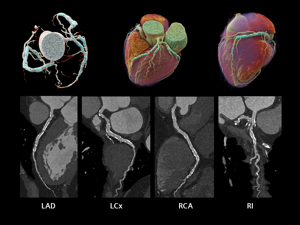
Fig 2. Top row: Cinematic volume-rendered technique (cVRT) images from CCTA, illustrating the three-dimensional coronary artery tree. Bottom row (from left to right): Curved planar reformats of the Left Anterior Descending (LAD), Left Circumflex (LCx), Right Coronary Artery (RCA), and Ramus Intermedius (RI), demonstrating the coronary lumen with diffuse, concentric, and heavily calcified plaques along the vessel walls. These findings reflect a severe and extensive calcific burden, consistent across all major coronary branches.
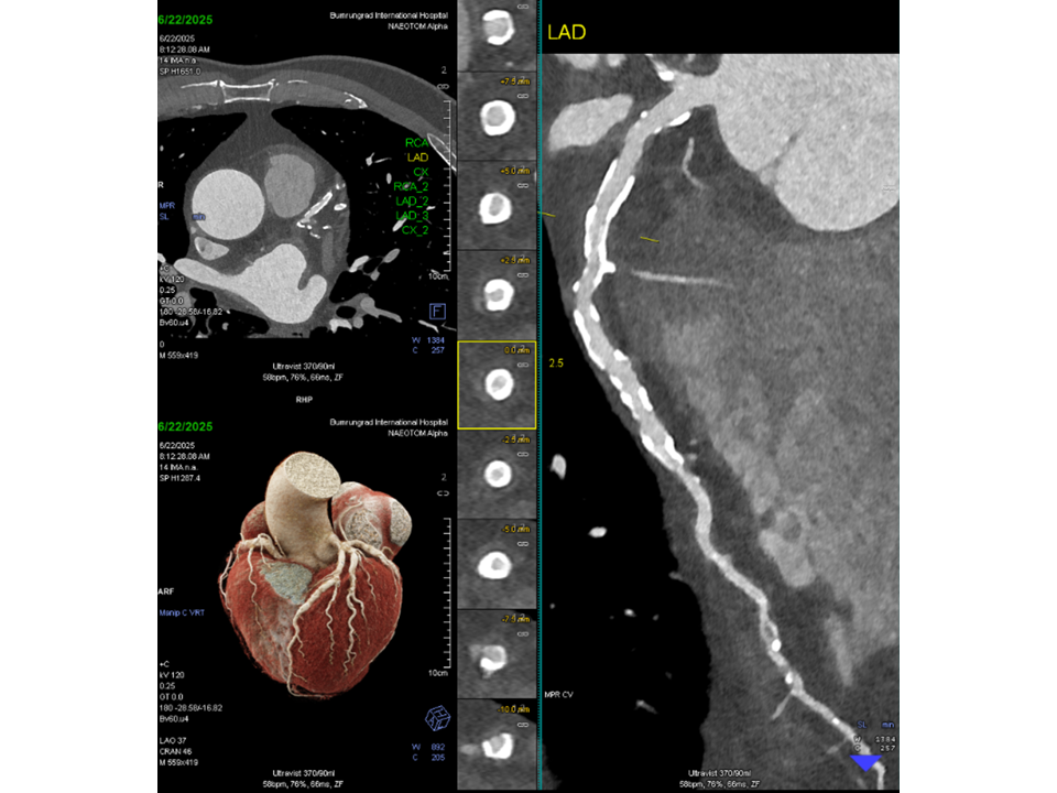
Fig. 3 Left panel: Axial and cVRT images from CCTA. Middle panel: Sequential axial slices through a coronary artery, illustrating cross-sectional views with dense calcified plaques embedded in the vessel wall of LAD. Right panel: Curved planar reformat of the LAD, showing diffuse and extensive calcification along the vessel wall with preserved lumen visualization, enabled by ultra high-resolution PCD-CT imaging.
Technical Advantages of PCD-CT
The NAEOTOM Alpha system, powered by PCD technology, offers several key advantages over traditional EID CT:
- Reduction of Blooming Artifacts: Dense calcifications often obscure the coronary lumen in conventional CT. PCD-CT minimizes blooming by reducing the influence of high-attenuation calcium on surrounding tissues, allowing clearer visualization of vessel patency.
- Enhanced Spatial Resolution: Submillimeter resolution at 0.2 mm enables more precise delineation of coronary anatomy, even in densely calcified segments.
- Lower Partial Volume Averaging: Smaller voxel dimensions reduce averaging across tissues, which helps prevent overestimation of plaque size and improves accuracy in lumen assessment.
- Improved Contrast-to-Noise Ratio (CNR) with Monoenergetic Imaging: Spectral reconstruction at optimal keV levels enhances contrast between iodine and calcium while suppressing image noise, improving diagnostic confidence in complex cases.
Discussion
This case highlights a pivotal advancement in cardiac imaging. The ability to confidently assess coronary arteries in a patient with a CAC score of 11,000 represents a breakthrough in non-invasive cardiovascular diagnostics. Previously, such cases would have been deemed non-diagnostic using EID CT, often necessitating invasive catheter angiography2.
The successful implementation of PCD-CT demonstrates the clinical value of this technology in real-world settings. It not only expands the diagnostic reach of CCTA but also improves patient care by reducing the need for invasive procedures in high-risk populations.
Conclusion
The integration of NAEOTOM Alpha with PCD marks a significant milestone in cardiac imaging. By pushing boundaries in the assessment of heavily calcified coronary arteries, this technology redefines the role of CCTA in patients with high CAC score. The case presented underscores the potential of PCD-CT to transform diagnostic pathways, offering precise, non-invasive evaluation even in the most challenging scenarios.

Written by
Dr. Achirawin Jirakamolchaisiri
Department of Cardiology,
Bumrungrad International Hospital, Bangkok, Thailand
Dr. Achirawin Jirakamolchaisiri is a leading interventional cardiologist at Bumrungrad International Hospital’s Heart Institute in Bangkok, with nearly 18 years of experience in catheter-based heart care. He earned his M.D. from Ramathibodi Hospital, completed comprehensive training in cardiology and internal medicine, and specializes in cutting-edge procedures, including PTCA, defect closures, pacemaker and CRT-D implantations, and RF ablation.
As Director of Heart Institute Operations, Dr. Achirawin champions a multidisciplinary approach to patient care, coordinating closely with cardiothoracic surgeons, geneticists, and rehab specialists. Fluent in English and Thai, he delivers patient-centered, minimally invasive heart treatments, making him a top choice for international patients seeking expert cardiac care in Thailand.