Cookies used for improved website experience

Siemens Healthineers and our partners use cookies and other similar technologies to operate the Siemens Healthineers websites and personalize content and ads. You may find out more about how we use cookies by clicking "Show details" or by referring to our Cookie Policy.
You may allow all cookies or select them individually. And you may change your consent and cookie preferences anytime by clicking on the "Review and change your consent" button on the Cookie Policy page.

syngo.via for Molecular Imaging animation of clinical images

syngo.via for MI

Reading as it should be in PET and SPECT

Today’s molecular imaging scanners are faster, more precise, and produce greater amounts of complex data than ever. Interpreting, managing, and sharing that data—across care teams, patients, devices, and time intervals—can present a host of challenges. 

syngo®.via for molecular imaging (MI) addresses these challenges by integrating everything you might require to read, interpret, report, and share your cases quickly and precisely on a single platform.
Features & Benefits

syngo.via’s unique combination of multiple smart technologies provides a level of automation that eliminates many of the manual pre-processing steps. With syngo.via, you can start reading right away and experience greater productivity.

Pre-fetching

Before opening a patient file, the prior examinations are automatically loaded onto syngo.via and are combined with the current exam in the SMART layout.

ALPHA technology

Proprietary ALPHA technology automatically correlates studies based on individual organ recognition and aligns them for more precise registration and easier evaluation.

Smart layout

Based on the type of scan performed, the appropriate application automatically opens in your preferred layout and displays the necessary tools.

Findings Navigator

See all previous findings within a case as soon as the case is displayed. Previous findings can be propagated to your current exam, allowing a quick comparison of patient studies.

Get the most out of your image, faster

syngo.via drives diagnostic clarity through a variety of unique MI algorithms that normalize quantification results and reduce variability across devices, patients, and time.

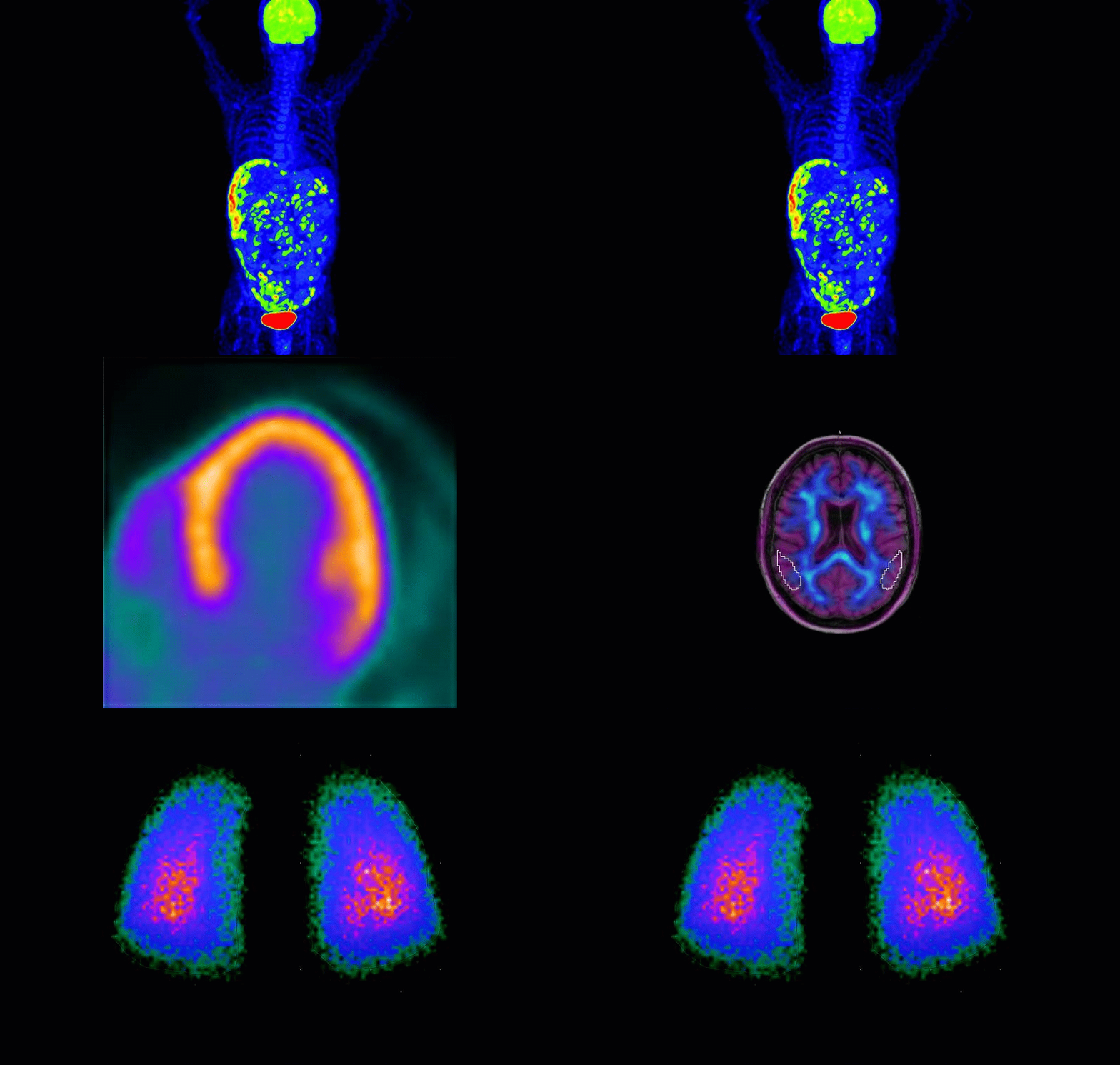
syngo.via for MI Oncolgoy - Key PET and SPECT features let you compare results from different time points and accurately track disease progression over time.

Oncology

Key PET and SPECT features let you compare results from different time points and accurately track disease progression over time.

syngo.via for MI Cardiology -Whether you prefer Corridor 4DM, Cedars Cardiac Suite or Siemens Healthineers applications, syngo.via allows you to read both SPECT and PET cardiac data on the same platform.

Cardiology

Whether you prefer Corridor 4DM, Cedars Cardiac Suite or Siemens Healthineers applications, syngo.via allows you to read both SPECT and PET cardiac data on the same platform.

syngo.via for MI Neurology - Our exclusive quantification algorithms provide quantitative guidance for the assessment of your diverse neurological indications.

Neurology

Our exclusive quantification algorithms provide quantitative guidance for the assessment of your diverse neurological indications.
syngo.via for MI Organ processing - Automated workflows let you perform advanced evaluation of general nuclear medicine datasets within the same interface.

Organ processing

Automated workflows let you perform advanced evaluation of general nuclear medicine datasets within the same interface.
syngo.via for MI - Collaborate across departments

From everyday reporting to mobile viewing and portable USB technology, syngo.via translates your data and findings into a concise report designed to enhance collaboration both online and in person.

Find your best fit—for today and tomorrow 

syngo.via’s high degree of flexibility and scalability supports a broad range of applications and users. It’s designed to meet your needs now and into the future.

syngo.via for MI multimodality applications


syngo.via multimodality applications

In addition to standard functionality, applications can be added to syngo.via any time.

syngo.via molecular imaging workplace


syngo.via molecular imaging workplace

Ideal for a stand-alone MI department or small private practice

syngo.via for MI multimodality workstation


syngo.via multimodality workstation

Ideal for a small hospital or stand-alone imaging center

syngo.via for MI multimodality server


syngo.via multimodality server

Ideal for a large hospital or practice with satellite branches

Clinical Use

syngo.via for MI - clinical use for oncology

Data courtesy of University of Tennessee Medical Center, Knoxville, Tennessee, USA. 

Our oncology application includes an array of features that help master increasing amounts of imaging data while supporting consistent measurement accuracy.

syngo.via for MI - clinical use for cardiology

Data courtesy of Manchester Royal Infirmary, Manchester, United Kingdom. 

Many physicians are working with both PET and SPECT cardiac data, and frequently use separate platforms to obtain a comprehensive view of a condition. syngo.via delivers the same consistent user experience for faster workflow, regardless of which modality or isotope utilized. Additional CT tools support your ability to interpret multimodality cardiology scans in a single platform.

syngo.via for MI - clinical use for neurology

Data courtesy of University of Tennessee Medical Center, Knoxville, Tennessee, USA. 

syngo.via provides quantitative guidance for the assessment of disease in both PET and SPECT cases, especially for borderline cases. Our dedicated neurological quantification algorithms help identify problems by comparing patient exams against a population database of normal, healthy brains, over time.

syngo.via for MI - clinical use organ processing

Data courtesy of University of Minnesota, Minneapolis, Minnesota, USA. 

syngo.via allows you to create and save automated workflows and process datasets using the same interface. Once a specific workflow is created, you can automate and replicate it — saving time and improving diagnostic confidence.

1