Clinical History
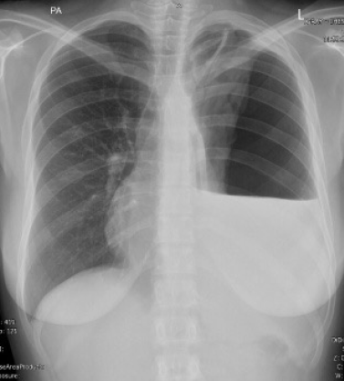
A 28-year-old female presented with a three-week history of persistent cough and progressively worsening dyspnoea. Initial chest radiography demonstrated a large left-sided hydropneumothorax with mediastinal shift, prompting emergent chest tube insertion. A post-procedural high-resolution computed tomography (HRCT) examination was subsequently performed using an ultra-high-resolution Dual-Source CT protocol to evaluate lung re-expansion and assess residual pathology.
Imaging Findings
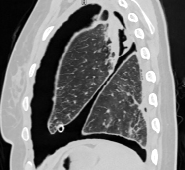
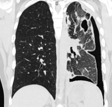
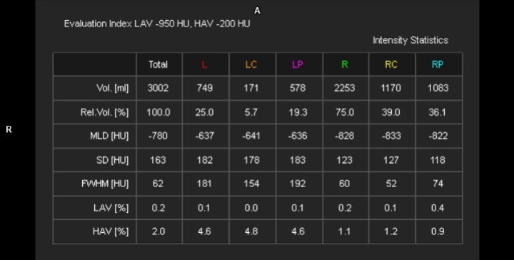
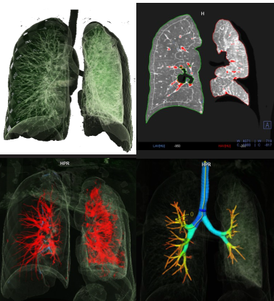
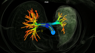
HRCT imaging, performed on the first Dual-Source Drive CT scanner in Kenya at The Nairobi Hospital, revealed a persistent large left pneumothorax causing significant rightward mediastinal displacement. Using syngo.via Pulmo 3D post-processing software, automated volumetric analysis demonstrated a marked asymmetry in lung volumes, with the left lung measuring 749 mL compared with 2,253 mL on the right. Passive atelectasis of the left lung was evident. Three-dimensional airway reconstruction showed significant compression of the left bronchial tree. Coronal reformatted images obtained following intervention demonstrated partial reduction of the pneumothorax with early signs of left lung re-expansion.
Discussion
Background
Tension pneumothorax is a life-threatening condition characterized by progressive accumulation of air within the pleural space, resulting in increased intrapleural pressure, mediastinal shift, and compromised cardiopulmonary function. Immediate recognition and prompt intervention are essential to prevent respiratory failure and hemodynamic collapse. Passive atelectasis frequently accompanies tension pneumothorax, occurring as a result of extrinsic compression of the lung parenchyma by intrapleural air or fluid.
Clinical Perspective
Patients with tension pneumothorax commonly present with dyspnoea, chest pain, hypoxia, and varying degrees of hemodynamic instability. Chest radiography typically serves as the initial diagnostic modality; however, it provides limited information regarding the extent of lung collapse and quantitative assessment of re-expansion. CT imaging plays a crucial role in confirming the diagnosis, identifying complications, and evaluating post-interventional outcomes. Quantitative lung volume assessment offers objective data for longitudinal monitoring and assists referring clinicians in evaluating therapeutic response.
Imaging Perspective
High-resolution CT offers superior anatomic detail for the evaluation of pneumothorax extent, lung collapse, and mediastinal displacement. The diagnostic value of this case is enhanced by the use of syngo.via Pulmo 3D software, which enables automated lung segmentation, volumetric quantification, and three-dimensional airway reconstruction. While standard HRCT provides qualitative assessment, Dual-Source CT combined with advanced post-processing tools enables rapid acquisition and objective quantification of lung volume loss. This approach improves diagnostic confidence and facilitates precise assessment of disease severity and treatment response.
Outcome
Management of tension pneumothorax primarily involves pleural decompression via chest tube insertion. Advanced imaging plays a critical role in determining the adequacy of lung re-expansion, identifying persistent atelectasis, and detecting complications such as bronchopleural fistula. Quantitative lung volume tracking establishes a baseline for assessing recovery and supports clinical decision-making regarding chest tube management and further intervention.
Take-Home Message
Advanced volumetric quantification using AI-driven post-processing software on Dual-Source CT platforms provides an objective and reproducible method for evaluating tension pneumothorax and monitoring lung re-expansion. The combination of Dual-Source CT acquisition with syngo.via Pulmo 3D enhances diagnostic precision beyond conventional qualitative assessment by delivering accurate lung volume measurements and detailed airway visualization. These capabilities support clinical decision-making, improve diagnostic confidence, and contribute to precision-based patient management in complex thoracic emergencies.
"The combination of Dual-Source CT acquisition with syngo.via Pulmo 3D enhances diagnostic precision beyond conventional qualitative assessment by delivering accurate lung volume measurements and detailed airway visualization."
The Nairobi Hospital
Nairobi, Kenya