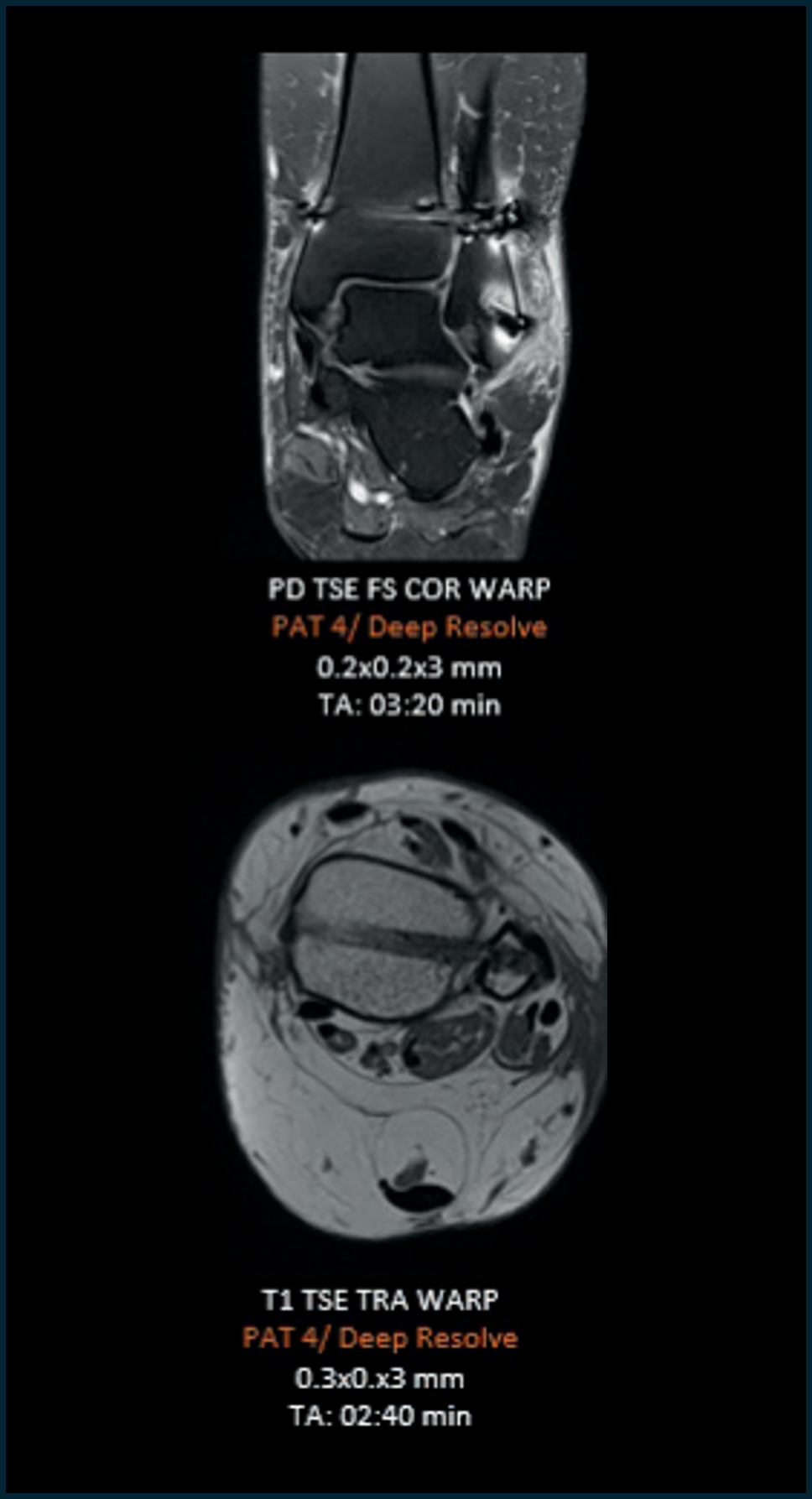
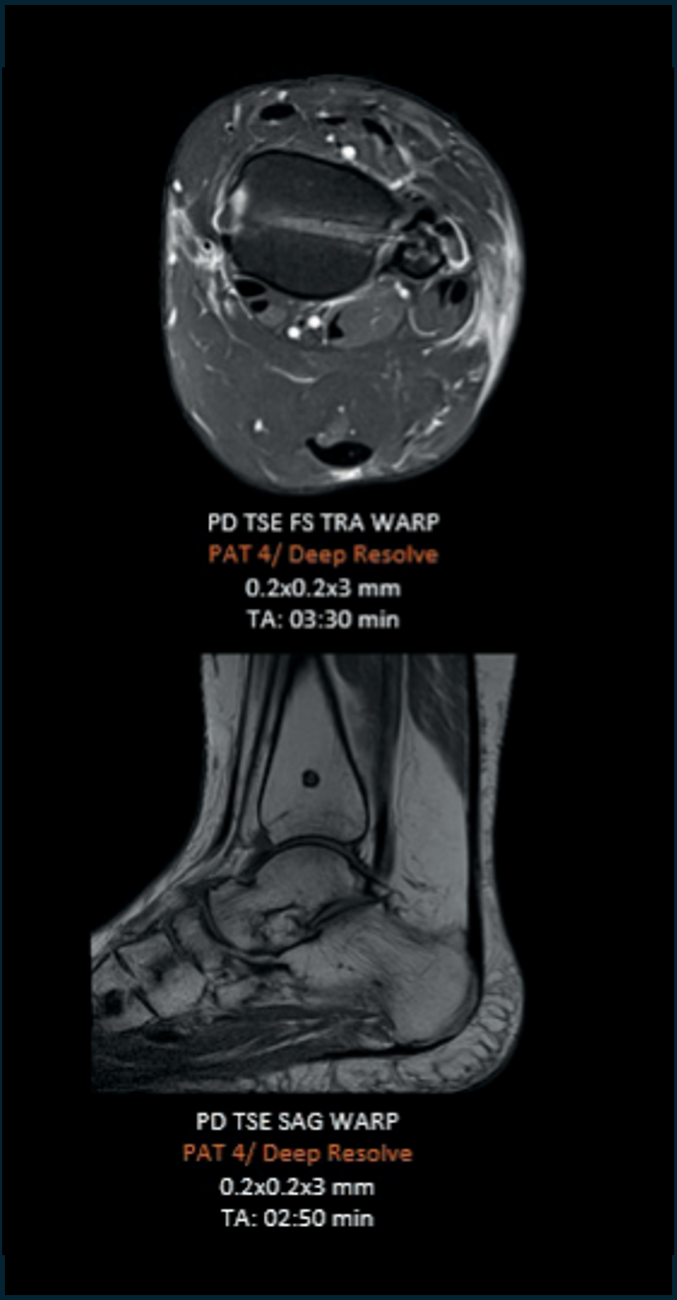
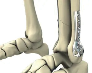
Metallic Implants: Imaging of Postoperative Changes
A growing number of patients with metallic implants present with postoperative or chronic clinical conditions that are most effectively evaluated using magnetic resonance imaging (MRI). MRI plays a critical role in the assessment of soft-tissue pathology, postoperative complications, infection, inflammation, and adjacent structural integrity. However, the presence of metallic implants—particularly those containing paramagnetic or ferromagnetic components—poses significant challenges due to susceptibility-related artifacts, signal distortion, and local field inhomogeneity, which can substantially limit diagnostic accuracy.
Imaging of Obese Patients
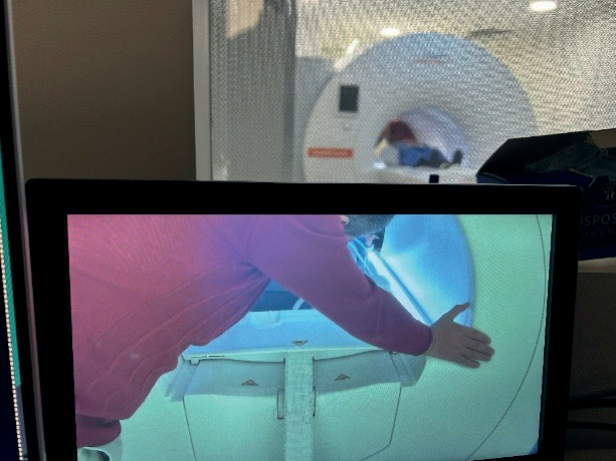
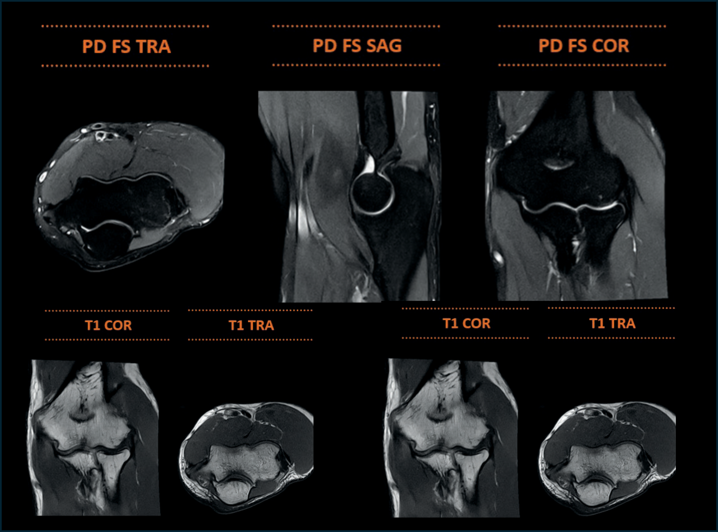
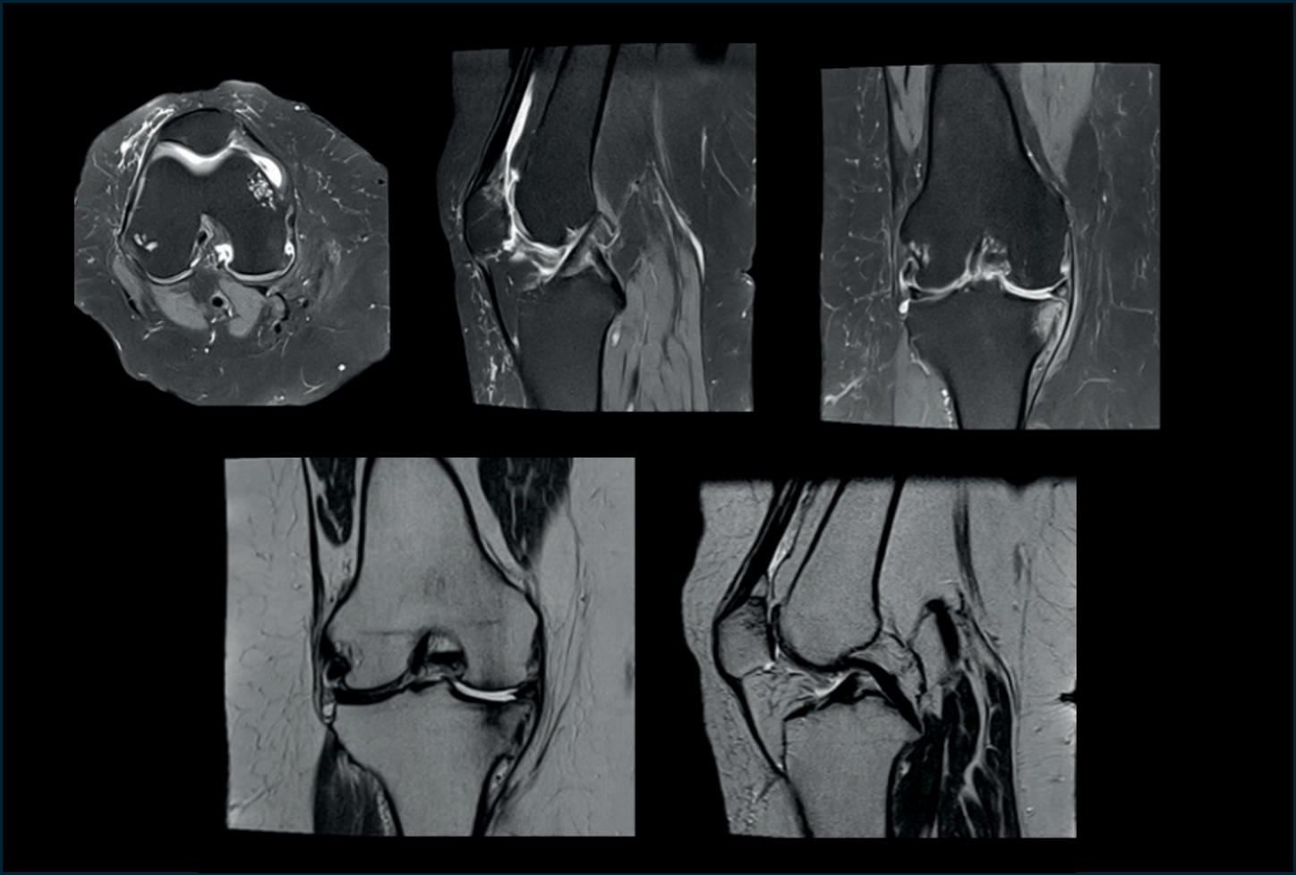
Obesity presents several challenges in magnetic resonance imaging (MRI), including limitations related to table weight capacity, bore diameter, patient positioning, claustrophobia, and difficulty maintaining immobility during image acquisition. These factors can adversely affect image quality and may result in incomplete examinations, motion artifacts, or exclusion of patients from MRI altogether. Addressing these challenges requires a combination of patient-centered system design, optimized imaging protocols, appropriate coil selection, and flexible patient positioning to ensure diagnostic-quality imaging.
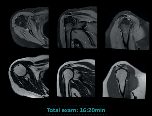
The MAGNETOM Free.Max system offers significant advantages for imaging obese patients through its open-bore design, which enhances both physical accessibility and psychological comfort. The wider bore allows patients to be positioned more comfortably and reduces the sensation of confinement, thereby improving patient cooperation and reducing motion-related artifacts. This design also facilitates flexible patient positioning, which is particularly beneficial for imaging off-center joints such as the shoulder and elbow, not only in obese patients but also across the general patient population.
Conclusion
Magnetic resonance imaging examinations can be challenging in specific patient populations, including individuals with obesity, metallic implants, and claustrophobia, often requiring specialized workflows and advanced imaging solutions to overcome technical and patient-related limitations. Addressing these challenges is essential to ensure diagnostic accuracy, examination completion, and equitable access to MRI.
The MAGNETOM Free.Max system enables a patient-centered imaging approach by combining low-field technology with an open-bore design and advanced image reconstruction capabilities. This platform facilitates successful MRI examinations in patients who are traditionally considered difficult to image, while maintaining diagnostic image quality and operational efficiency. Clinical experience demonstrates that the use of MAGNETOM Free.Max expands MRI accessibility and supports inclusive, high-quality imaging across diverse patient groups, underscoring its value in contemporary radiologic practice.
"The MAGNETOM Free.Max system enables a patient-centered imaging approach by combining low-field technology with an open-bore design and advanced image reconstruction capabilities."