
Clinical Case Study 1
Free-Breathing Lung Imaging with Turbo Flash
Patient History and Objective
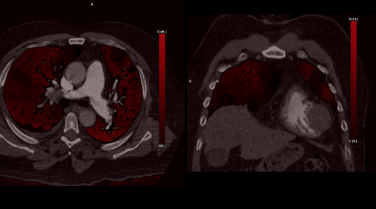
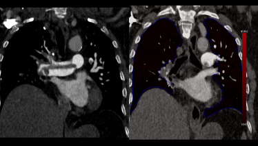
A 65-year-old male patient weighing 120 kg presented with breathing difficulties and recent-onset chest pain. The patient had no prior history of chest pain or related symptoms. Due to clinical suspicion of pulmonary embolism (PE), a CT pulmonary angiography (CTPA) study was recommended. Given the patient’s inability to hold their breath, a free-breathing imaging protocol was deemed essential to ensure diagnostic accuracy while addressing challenges related to his body habitus.
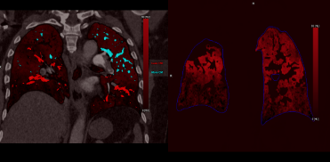
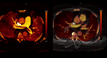
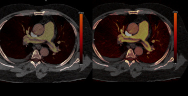
This study aimed to evaluate the feasibility and clinical impact of free-breathing lung imaging using the Turbo acquisition mode in PCCT, focusing on dose reduction, contrast optimization, and functional perfusion analysis.
Clinical Case Study 2
Photon-Counting CT Imaging: Redefining Diagnostic Standards with Low-Dose Cardiac Imaging in Single-Beat Turbo Flash Mode with Full Spectral Capabilities
Photon-counting CT (PCCT) with Turbo Flash mode represents a groundbreaking advancement in cardiac imaging, combining ultra-high spatial and temporal resolution with intrinsic spectral capabilities—all at remarkably low radiation doses.
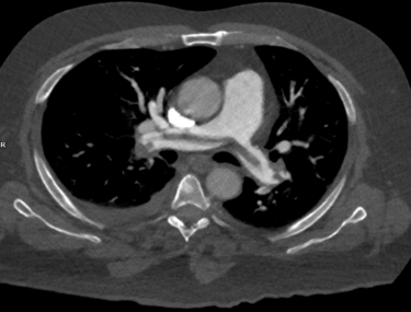
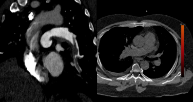
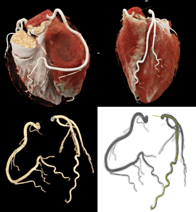
This study employed Turbo Flash single-beat acquisition to achieve a slice thickness of 0.4 mm, enabling exceptionally detailed visualization of cardiac anatomy. The ultra-fast imaging facilitated by Turbo Flash mode offers temporal resolution far superior to conventional CT systems, making it possible to perform single-beat cardiac imaging with comprehensive spectral data acquisition. Spectral analysis, and functional assessment.
Patient History and Objective:
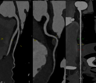
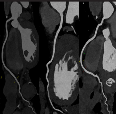
A 65-year-old male with a history of medically managed coronary artery disease, previously treated with stent placement, presented for a follow-up evaluation. The objective of the study was to assess stent patency and evaluate the surrounding vasculature for signs of restenosis or other abnormalities.
Given the patient’s clinical history, high-resolution imaging was deemed essential to provide detailed visualization of the stent and adjacent vascular structures, facilitating a comprehensive diagnostic assessment and guiding ongoing management.
Conclusion
In conclusion, photon-counting CT with ultra-low-dose, high-pitch Turbo Flash single-beat imaging represents a transformative advancement in cardiac diagnostics. This innovative approach delivers unmatched spatial and temporal resolution with very low-dose delivery, enabling precise visualization of cardiac structures and functional assessment in a single heartbeat. The capability to acquire high-quality, motion-free images with a sub-millisievert radiation dose ensures patient safety without compromising diagnostic accuracy.
Intrinsic spectral imaging further enhances the protocol by providing detailed insights into myocardial perfusion, plaque characterization, and vascular health. By combining ultra-fast acquisition with comprehensive spectral data, photon-counting CT establishes a new gold standard in cardiac imaging, offering unprecedented precision, efficiency, and safety for both routine and complex clinical cases.
"Photon-counting CT with ultra-low-dose, high-pitch Turbo Flash single-beat imaging represents a transformative advancement in cardiac diagnostics."

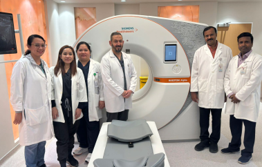
Al Ahli hospital, Qatar
"Intrinsic spectral imaging further enhances the protocol by providing detailed insights into myocardial perfusion, plaque characterization, and vascular health."

Al Ahli hospital, Qatar