Triple-rule-out evaluation of acute chest pain using single source photon-counting CT

Lili Száraz, MD; Sámuel Beke, MD; Kristóf Nagy, MD; Barnabás Baksa, MD; Péter Hegedűs, MD, PhD; Prof. Pál Maurovich-Horvat, MD, PhD
Medical Imaging Center, Semmelweis University, Budapest, Hungary

2026-01-29
A 50-year-old male patient presented to the emergency department (ED) with massive hemoptysis, dyspnea, and chest pain. The patient’s medical history included multiple cardiac risk factors and adverse events, including hypertension, chronic obstructive pulmonary disease, previous myocardial infarction, and mitral valvuloplasty. Notable physical exam findings included tachypnea, spastic respiratory sounds with left-sided crepitations, and diaphoretic skin. Laboratory findings revealed elevated inflammatory markers, high-sensitivity Troponin T, and D-dimer levels. Given the high suspicion of pulmonary embolism (PE), a targeted chest CT angiography (CTA) was performed, using ECG-gated helical acquisition with standard-resolution scan mode, to confirm the PE and evaluate the bronchial arteries, guiding further potential embolization therapy.
The multiphase chest CTA confirmed a left-sided PE complicated by infarct pneumonia (see Figure 2). Thrombolytic therapy was not indicated, as determined in consultation with the intensive care unit. Likewise, bronchial artery embolization was considered unnecessary, since the hemoptysis was attributed to the severe infarct pneumonia. Empirical antibiotic therapy was initiated and later escalated to a combination of piperacillin–tazobactam and metronidazole. Due to significant hemoptysis, anticoagulation was administered at a reduced dose; nevertheless, anti-Xa levels remained within the therapeutic range. The chest CTA additionally revealed a left hilar mass infiltrating the pulmonary branches of the left lower lobe (see Figure 2), as well as a smaller, round lesion closely adherent to the pericardium at the base of the right lung. On a one-month follow-up scan, the left-sided mass had resolved, while the right-sided benign-appearing lesion remained unchanged. On a three-month follow-up scan, no malignancy was found, the atelectasis and consolidations showed signs of regression, however the segmental and subsegmental emboli were still visible.
A cVRT image of the heart and curved multiplanar reconstructions of the three coronary arteries, namely the right coronary artery, the left main, and the left anterior descending. A complete myocardial bridge is noted in the mid-segment of the left anterior descending artery. The coronary arteries are clearly visualized despite a high and irregular heart rate ranging from 86 to 158 beats per minute, as documented in the ECG file.
Courtesy of Semmelweis University, Medical Imaging Center, Budapest, Hungary

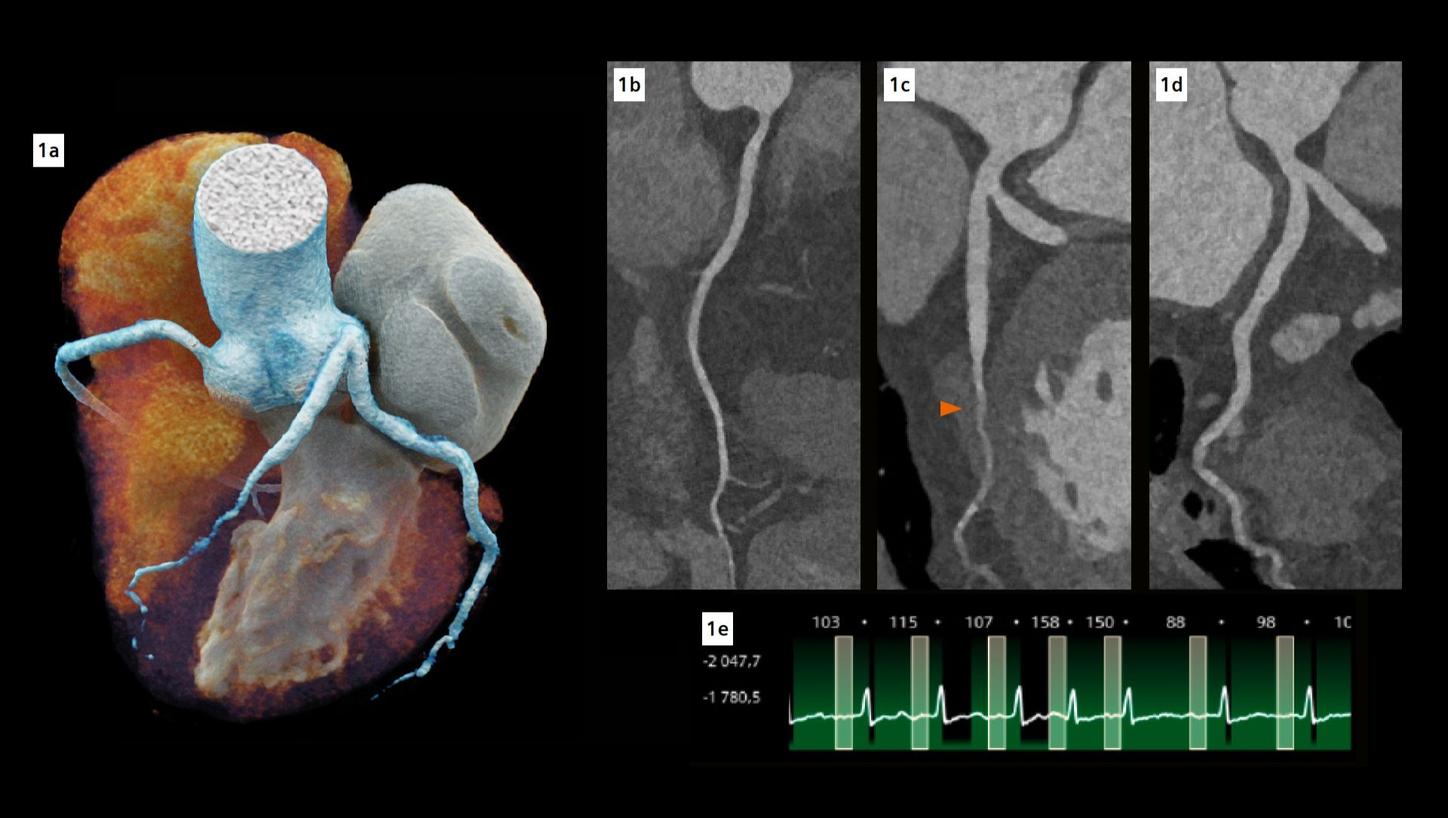
Fig. 1: Cinematic volume rendering technique (cVRT) image of the heart (a) and curved multiplanar reconstructions of the coronary arteries (b-d). (b) represents the right coronary artery, (c) represents the left main and left anterior descending (LAD) coronary artery, while (d) represents the left main and left circumflex coronary artery. The arrow indicates a complete myocardial bridge on the middle LAD. Note the excellent visualization of the coronary arteries despite high and irregular heart rate (e).

A filling in the lower part of the left pulmonary artery is shown on a coronal MIP image of the pulmonary vasculature and on a cross-sectional view of a VRT reconstruction of the pulmonary vessel arborization. Finally, an oblique view of a VRT reconstruction of the thoracic vessels shows the embolus highlighted in blue, and a cloud-like visualization of the consolidations in the left lung.
Courtesy of Semmelweis University, Medical Imaging Center, Budapest, Hungary

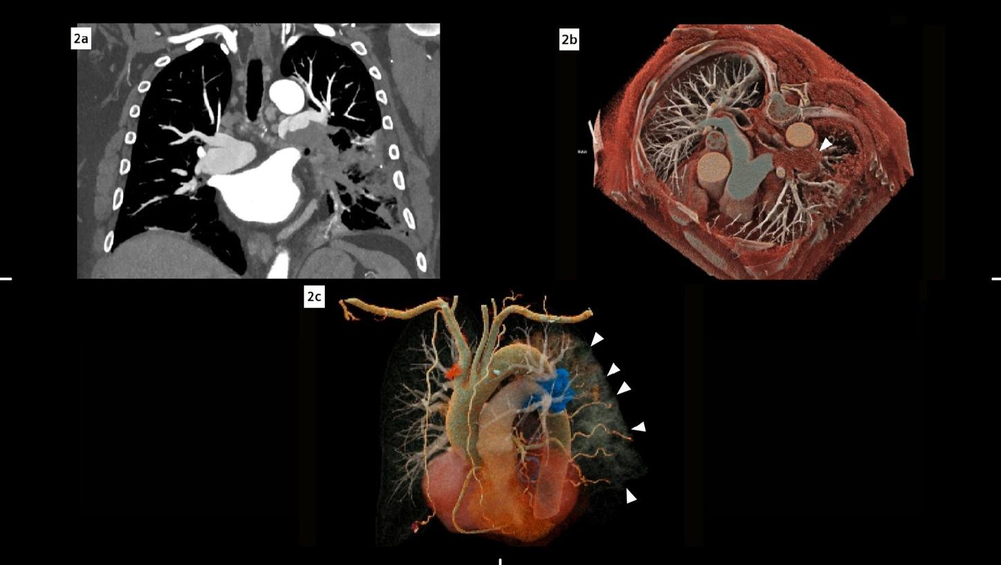
Fig. 2: Coronal maximum intensity projection (MIP) image of the pulmonary vasculature (a) demonstrates a filling defect in the lower part of the left pulmonary artery. Volume rendering technique (VRT) reconstructions (b, c) depict the pulmonary vessel arborization: In (b), a cross-sectional view, the filling defect is visible in the left pulmonary artery (arrow). In (c), an oblique view, the embolus is highlighted in blue, while the consolidations in the lung can also be appreciated (small arrows).

While the ability of triple-rule-out-CT (TRO-CT) to rule out disorders in all three vascular beds (pulmonary embolism, PE; aortic dissection, AD; and coronary artery disease, CAD) has been demonstrated [1], a gap remains in the literature regarding the clinical impact of TRO-CT. Araoz P.A. et al. found that, based on accepted clinical risk scores, 65% of their patient cohort warranted work-up for at least two of either PE, AD or CAD suggesting that a large subset of patients would benefit from a comprehensive test like TRO-CT [2], even though the mean radiation dose (23.8 ± 12 mSv; range: 3.7–84.1 mSv) and contrast media usage (144.6 ± 31.2 mL; range: 54-360 mL) varied widely. On the other hand, Takx et al. found that TRO-CT may reduce the length of stay and costs in ED in appropriate patients, but no difference in the diagnosis of PE, AD, or CAD was observed between TRO-CT versus standard-of-care strategies [3]. Currently, no major US or EU guidelines recommend routine TRO-CT as the initial ED test for undifferentiated acute chest pain, due to the limited contemporary comparative evidence. This case report serves as a reference, suggesting that PCD-CT may help revive the TRO-CT paradigm. 

In this case, the targeted chest CTA enabled the use of a TRO-CT equivalent methodology. Therefore, the entire thoracic aorta was included in the scan range and timing was set to achieve optimal enhancement in the aorta and pulmonary arteries. Due to the high spatial resolution of the photon-counting detector, the coronary arteries could also be accurately evaluated. 

The fine spatial detail (0.4 mm slice thickness) enabled an accurate evaluation of the coronary arteries to rule out severe CAD (as part of the differential diagnosis of chest pain) (see Figure 1). The use of advanced image reconstruction algorithms, such as ZeeFree, combined with the preview series function, resulted in only minor motion artifacts on the acquired scans, despite the high heart rate variability (Minimum: 86, Maximum: 158, average: 110). Detector-based quantum spectral imaging capabilities (lung analysis reconstruction) made it impressively fast to detect lung perfusion deficits caused by the PE and aided further therapy decision-making by down-grading disease severity. A fair dose length product (636 mGy*cm) was achieved during the chest CTA, utilizing the NAEOTOM Alpha.Prime CT scanner with an optimized protocol, serving as a true “one-stop-shop” TRO-CT examination.

Scanner

Scan area

Chest

Scan mode

QuantumPlus

Scan length

325.95 mm

Scan direction

Cranio-caudal

Scan time

4.881 s

Tube voltage

140 kV

Effective mAs

281.15 mAs

IQ level

64

Dose modulation

CARE Dose4D

CTDIvol

22.4 mGy

DLP

636 mGy*cm

Rotation time

0.25 s

Pitch

0.29

Slice collimation

0.4 mm

Slice width

57.60 mm

Reconstruction increment

0.2 mm (cardiac), 1 mm (lung)

Reconstruction kernel

Bv44 (cardiac), Qr40 (lung)

keV level

70 (cardiac), 55 (lung)

Spectral reconstruction

VMI, Lung Analysis, VNC, Iodine

Heart rate

110 (range: 86–158)

Contrast

350 mg/mL

Volume

80 mL + 50 mL saline

Flow rate

4.5 mL/s

Start delay

Bolus tracking triggered at 200 HU in the ascending aorta

[1]
[2]
[3]