Introduction
Mammary carcinoma is the most common malignant tumor in women, and it is the leading cause of mortality, with an incidence of >1,000,000 cases occurring worldwide annually. Invasive ductal carcinoma (IDC), also known as infiltrating ductal carcinoma, is the most common type of mammary carcinoma; 80% of all mammary carcinoma are invasive ductal carcinomas.
Invasive indicates that the cancer has invaded or spread to the surrounding breast tissues and ductal carcinoma refers to the cancer growing in a milk duct that has invaded the fibrous or fatty tissue of the breast outside of the duct.
New technologies that influence our mammography imaging practice
When performing Contrast Enhanced Dual Energy Mammography on a standard mammography system, the high-energy (HE) and low-energy (LE) images must be acquired successively, With the introduction of the MAMMOMAT Revelation, Siemens Healthineers has implemented its Titanium Contrast-Enhanced Mammography (TiCEM) CEDEM application. TiCEM aims at improving diagnostic accuracy in the detection and characterization of breast tumors, by incorporating functional information.
The clinical workflow for a TiCEM examination starts with the injection of the iodinated contrast agent by means of a power injector. At the time of injection, the breast is not (yet) compressed, to allow for normal tissue perfusion and unhindered inflow of the contrast agent into the breast. The dosage of the contrast agent is typically weight-dependent and varies between institutions. After a waiting time of approximately 2 minutes, the woman is positioned at the MAMMOMAT Revelation and the breast is compressed. Then, a low-energy (LE) and a high-energy (HE) image are acquired successively and an Insight CEM image, a recombined image of that view is calculated. These steps are then repeated for each additional view, without the need to perform a new contrast agent injection. The time window for performing multiple views with a single contrast agent injection lasts up to 10 minutes, although the views should be acquired without any unnecessary delays. The order in which the views are acquired seems to be of little clinical significance and does not appear to affect image quality. Care should be taken when handling the contrast agent to avoid contamination of the detector or the skin with pure contrast agent, as this might mimic calcifications or result in artifacts.
In breast care, Tomosynthesis technology – also known as three-dimensional (3D) mammography revolutionizes diagnostic mammography. Tomosynthesis technology is slated to soon take over full field digital mammography (FFDM) by means of improved diagnostics, better image quality, reduced recall rates, workflow etc. Tomosynthesis technology creates an impact in almost every aspect of breast imaging from diagnostic breast cancer screening to interventions. It helps us to reduce the recall rate and false positive rates, irrespective of a women’s age or breast density.
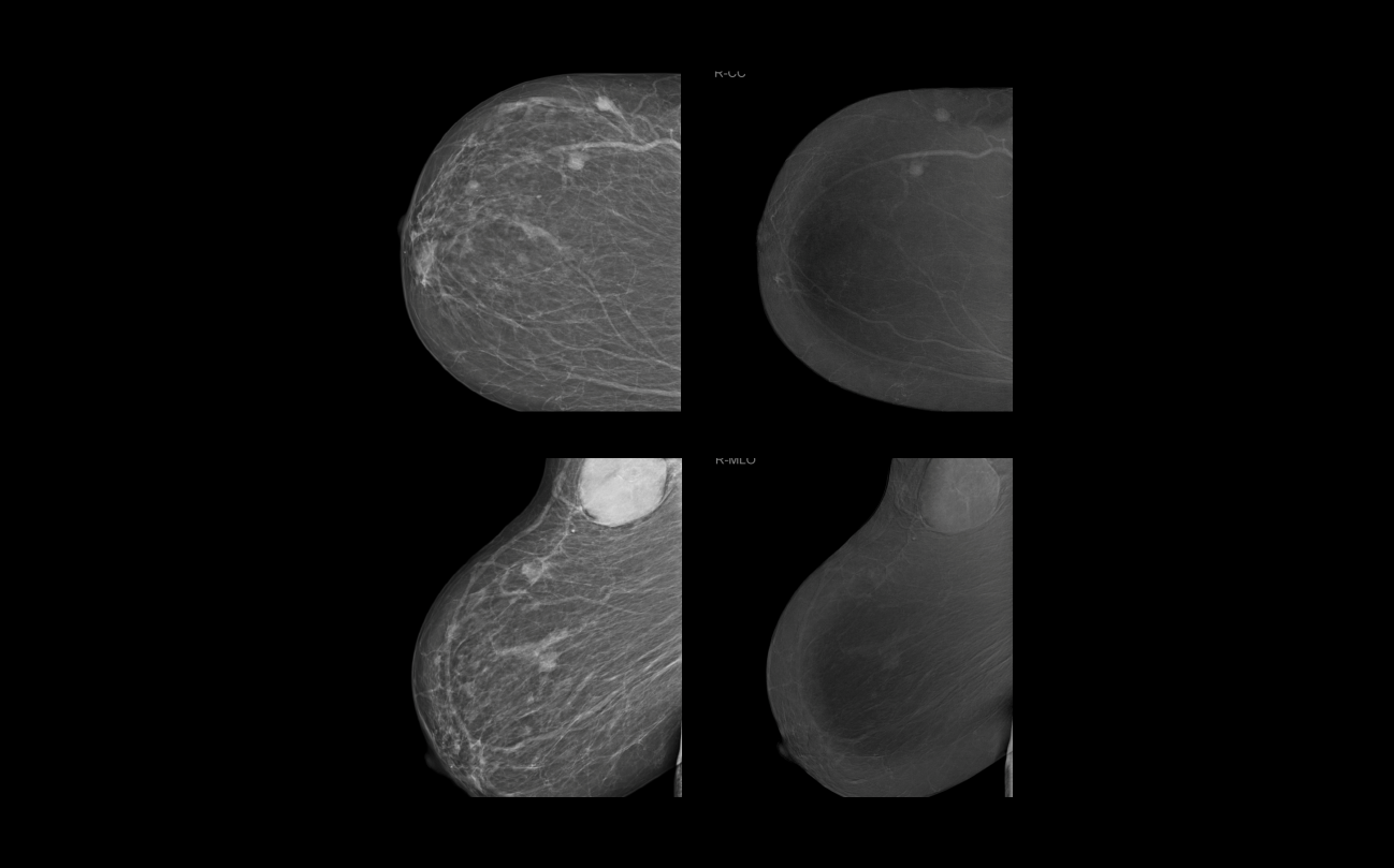
TiCEM with 50° wide-angle HD helps to change clinical management from surgery to therapy in IDC
Clinical findings
The patient is a 55-year-old female patient with a known history of carcinoma. The mediolateral oblique (MLO) standard mammographic views delineate the IDC from the previous examination. The initial plan was to do a conservative surgery. Further imaging with CEDEM prospective assessment revealed multiple non-palpable small nodules and enhancement in high energy contrast imaging. Late enhancement images outlined the axillary masses with adjacent lymph nodes. IDC was confirmed by the biopsy and other advanced imaging techniques which led to the decision of other clinical management in place of the surgical plan.
Conclusion
Siemens Healthineers pioneered the 50° wide-angle HD Breast Tomosynthesis, reaching the highest depth resolution on the market (3.5 times higher depth resolution compared to narrow angle systems) and Titanium Contrast Enhanced Mammography (TiCEM) with its unique HE spectrum and an optimized titanium filter, which reduces X-ray tube load to enable seamless examinations. TiCEM delivers additional diagnostic information for more confident decision-making and helps to detect or rule out lesions. Being an integrated functionality of the Siemens Healthineers MAMMOMAT Revelation, TiCEM can help reduce scheduling conflicts and workload on other modalities making it a cost-effective alternative to breast MRI. Finally, guidelines are needed to achieve international standards in acquisition techniques and image interpretation.
Contact

Contact

Head of Radiology Dept., Women’s Health Unit,
Assiut University Hospitals, Assiut University, Assiut, Egypt