In breast care, Tomosynthesis technology – also as known as three-dimensional (3D) mammography revolutionizes diagnostic mammography. Tomosynthesis is much superior to basic digital mammography in several aspects which include improved diagnostics, better image quality, work-flow etc and therefore Tomosynthesis technology creates an impact in almost every facet of breast imaging that includes diagnostic breast cancer screening to interventions. The technology helps reduce the recall rate as well as false positive rates, irrespective of a women’s age or breast density.
The R&D and architecture that goes into a tomosynthesis system requires thoughtful consideration of an enormous number of parameters that leads to a meaningful clinical solution. The Siemens Healthineers MAMMOMAT Revelation, a product of many years of research and clinical experience is the first and only wide angle 50-degree tomosynthesis mammography system in the industry as of today.
The Siemens Healthineers MAMMOMAT Revelation is made first and foremost to ensure the comfort of the patient. Ease of use and soft compression technology helps clinicians, achieve ultimate patient comfort and relaxation. The technology which includes highest depth resolution and 50° HD Breast Tomosynthesis Leverage precision in high quality diagnostic mammography imaging, gives clinicians a more clear and confident diagnosis even in complex clinical scenarios.
Clinical Evidence – Tomosynthesis - Case 1
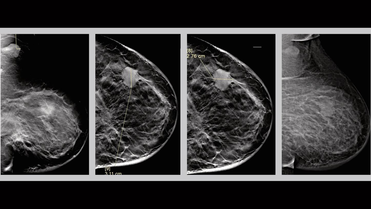
History
Patient is 63 years old presented for a bilateral diagnostic mammography. The results showed palpable left breast mass and pathologically enlarged left axillary lymph nodes.
Tomosynthesis Mammography Findings
Finding 1
There is a high density, irregularly bordered mass measuring 3 cm with speculated margins seen in the middle third of the left breast upper outer quadrant (1 o’clock position), located 8 cm away from the nipple. Irregular mass correlates to the clinically palpable area. There are indeterminate calcifications with grouped distribution within the mass.
Finding 2
There is an area of architectural distortion with associated mass, best visualized through tomosynthesis images. It is seen at the posterior aspect of the left breast, lower inner quadrant, measuring 3 cm, and located 10 cm from the left nipple. There are fine linear branching calcifications within this area also best visualized by tomosynthesis images. Finding 2 is 8 cm posterior to finding 1 (the index lesion). Localized overlying skin thickening is noted at the lower central part of the left breast.
Finding 3
Pathologically enlarged left axillary lymph nodes. Cortical thickness reaching 0.6 cm.
Clinical Evidence – Tomosynthesis - Case 2

History
Patient is 65 years old and is seen for palpable left breast lump. No personal or family history of breast cancer or any other cancer.
Mammography Findings
The following mammographic tomosynthesis 3D views were obtained as bilateral digital craniocaudal and bilateral digital mediolateral oblique.
Finding 1
There is a high density, irregular mass measuring 3 cm with speculated margins seen in the middle third of the left breast (2 o’clock position) located 6.5 cm from the nipple. Irregular mass correlates to palpable area.
Finding 2
There is a suspicious left axillary lymph node with non-uniform thickened cortex reaching 0.6 cm.
Finding 3
Few small circumscribed masses in the right breast. No suspicious microcalcifications or architectural distortion evident in the right breast.
Impression
BIRADS Assessment Category 5: Malignant Finding
Clinical Evidence – Tomosynthesis - Case 3
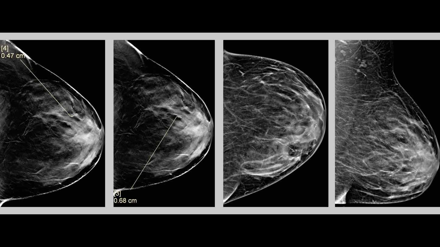
History
Patient is 46 years old and is seen for screening mammography. No personal or family history of breast cancer or any other cancer.
Mammography Findings
Finding 1
There are three circumscribed equal density masses noted on tomosynthesis images, in the anterior aspect of the left breast, at the upper outer quadrant (2 o’clock position), about 4 cm from the nipple. They measured about 0.6 cm.
Impression
BIRADS Assessment Category 2: Benign finding

Dr. Hanan Gewefel, MD. EDBI,
Founder & CEO WAFI Center
References
1. Chan H-P, Wei J, Sahiner B, et al. Computer-aided Detection System for Breast Masses on Digital Tomosynthesis Mammograms: Preliminary Experience. Radiology. 2005;237:1075–1080.[PMC free article] [PubMed]
2. Singh S, Tourassi GD, Baker JA, Samei E, Lo JY. Automated breast mass detection in 3D reconstructed tomosynthesis volumes: a featureless approach. Medical Physics. 2008;35:3626–3636. [PMC free article][PubMed]
3. Reiser I, Nishikawa RM, Edwards AV, et al. Automated detection of microcalcification clusters for digital breast tomosynthesis using projection data only: a preliminary study. Medical Physics. 2008;35:1486–1493. [PMC free article] [PubMed]
3. Reiser I, Nishikawa RM, Giger ML, et al. Computerized mass detection for digital breast tomosynthesis directly from the projection images. Medical Physics. 2006;33:482–491. [PubMed]
4. Svane G, Azavedo E, Lindman K et al. Clinical Experience of photon counting breast tomosynthesis: a comparison with traditional mammography. Acta radiol 2011: 52 92): 134-42.
5. Spangler M, Zuley M & Sumkin J. Detection and Classifications of Calcifications on Digital Breast Tomosythesis and 2D Digital Mammography: A Comparison. Am J Roentgenol 2011: 196: 320-324.
6. Wallis MG, Moa E & Zanca F. Two-view and single-view tomosynthesis versus full-field digital mammography: high-resolution X-ray imaging observer study. Radiology 201: 262 (3): 788-96.