
For routine MR examinations, reimbursements are falling and referrals are increasing due to changing demographics. Powered by artificial intelligence GO Technologies help you accelerate the entire workflow from patient positioning to result distribution. With the holistic set of intuitive workflow automations it is possible to reduce the average MRI exam slot from 30 to 20 minutes. This allows you to schedule one more patient per hour1.

GO Technologies
Power your MRI workflow
Patient preparation with Select&GO
With BioMatrix Select&GO laser positioning becomes obsolete. It lets operators use artificial intelligence to automatically position patients up to 30%1 faster and avoid repositioning delays. The user simply selects the region or organ to be scanned with one touch on the display and the patient is precisely positioned.
Image acquisition with DotGO

Reduce inter-operator variability and get consistent image results with DotGO. Our unique MRI exam software streamlines your MRI workflow and standardizes your clinical quality. DotGO consists of two components: The Dot Cockpit and the Dot engines. The Dot Cockpit is your central exam user interface for intuitive protocol configuration and management. The Dot engines provide you dedicated workflow packages for efficient and guided scanning.
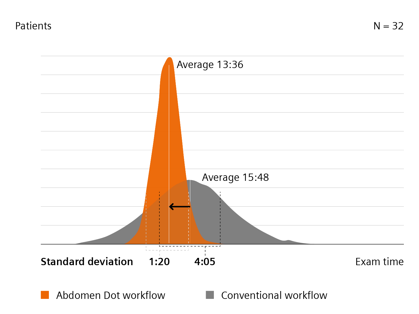
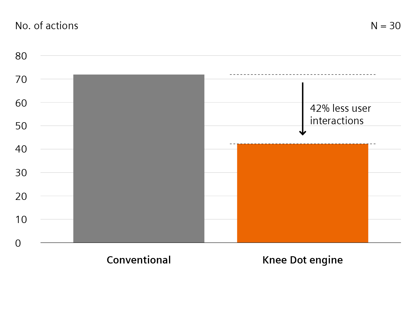
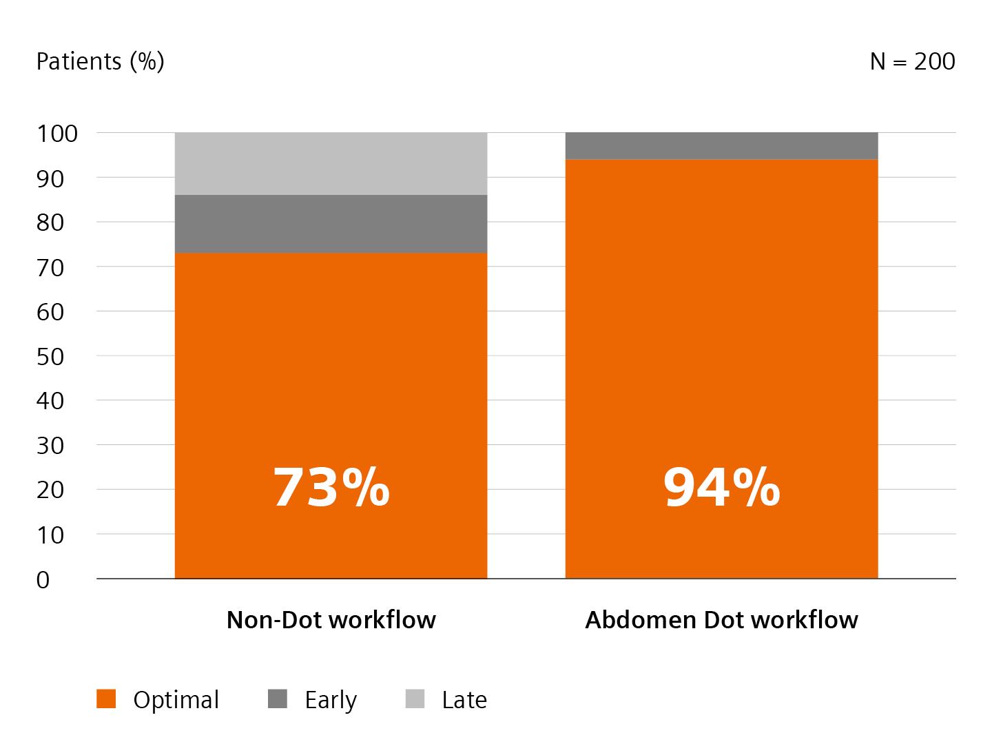
Image reconstruction with Recon&GO

Save valuable time and reduce workflow steps with inline reconstruction. Recon&GO automates reconstruction tasks with zero clicks. For example, inline composing of whole spine exams and vertebrae labeling in all planes can be performed without any user interaction.
Result distribution with MR View&GO

Distribute your results in one user interface without the need to search for patient data in the patient browser. MR View&GO automatically displays currently scanned data for quality assurance and viewing. It provides intuitive tools that manage and distribute results efficiently. In addition advanced applications such as generating computed high b-value images or 3D reconstructions of the plexus can be performed directly at the scanner, reducing the workload for radiologists.
Avez-vous jugé cette information utile?
1
Data on file.
2
Zhongshang Hospital Fudan University, Fudan, CN, Abdomen Dot Engine Workflow Study.
3
Knee Dot Engine Workflow Study: Prof. Bremer, St. Franziskus Hospital, Münster, Germany, 2014.
4
Martin, Diego R., “Optimization of Single Injection Liver Arterial Phase Gadolinium Enhanced MRI Using Bolus Track Real-Time Imaging.”, Journal of Magnetic Resonance Imaging; 33:110-118 (2011).
5
“Cardiac Dot Engine: Significant Time Reduction at Cardiac Magnetic Resonance Imaging”, MAGNETOM Flash.
6
“fit-Upgrade: A Success Story”; MAGNETOM Flash ISMRM edition, 2015.