NeurologyShaping the future of neurovascular care with CT and AI
Our mission is to help neurology patients by supporting their diagnosis and treatment. We are committed to providing you with fast and reliable CT imaging to make informed treatment decisions. They are, of course, particularly important in stroke, where time is brain.
We firmly believe that innovation in computed tomography can create great momentum in neuroradiology and neurology. You think ahead in advancing neurology care. We innovate ahead with technologies that support you throughout the entire patient pathway. Discover how integrated neurology innovations by Siemens Healthineers can help you care for your neurological patients.
Highlights & Innovations
Innovating in neuroradiology with the NAEOTOM Alpha class
Our breakthrough photon-counting technology enables a profound clinical impact beyond the reach of conventional CT. The NAEOTOM Alpha class lets you visualize anatomy and characterize materials in high detail while keeping dose low.
Transforming critical care workflows with SOMATOM On.site
Scanning patients directly in the ICU helps avoid complications and improves efficiency. Prof. Johan Wasselius, MD, explains how point‑of‑care imaging with SOMATOM On.site has enhanced critical care workflows at Skåne University Hospital in Lund, Sweden.
Clinical Applications
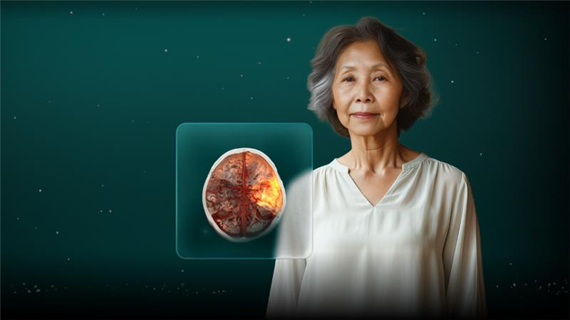
When every minute matters imaging empowered by our CT Neuro AI applications make the difference.
Learn how we can speed up stroke imaging and reading and make a difference to stroke survivors like Jenny.
Clinical Cases
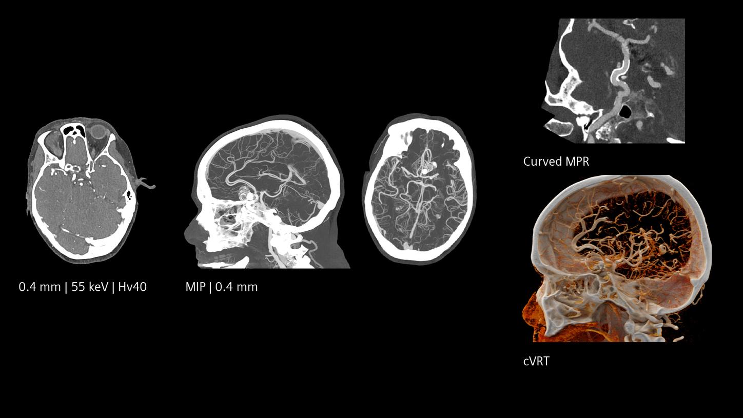
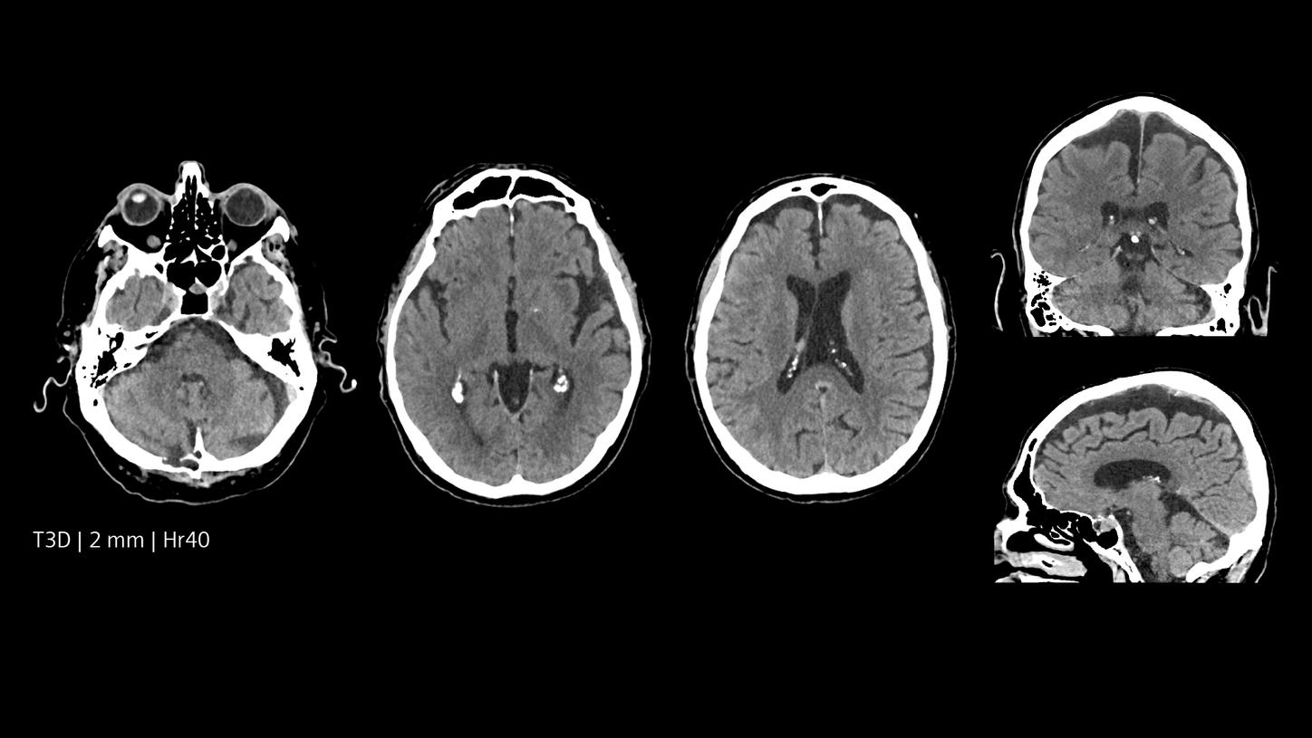
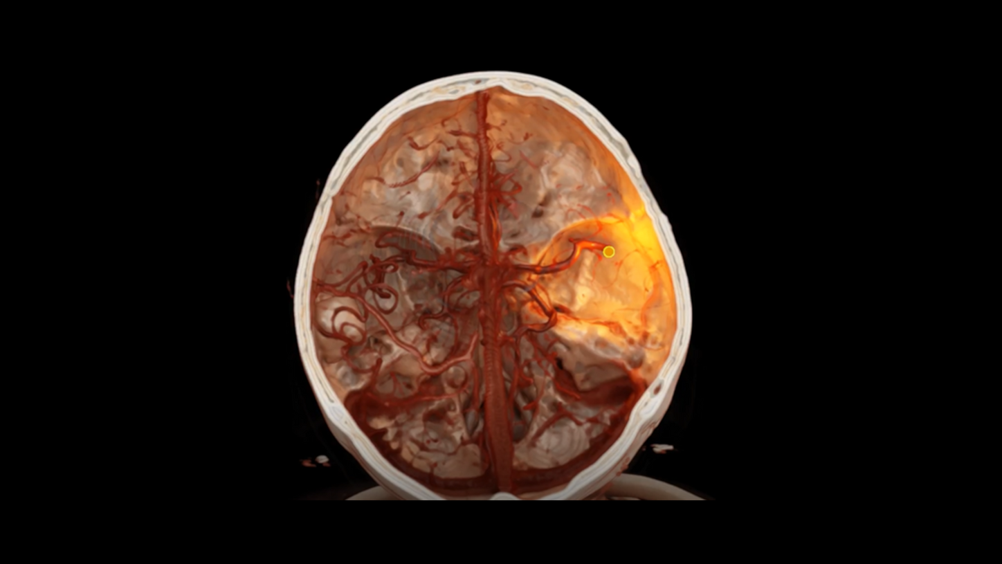
CT angiography of the cerebral arteries
NAEOTOM Alpha | CTDIvol 8.5 mGy
Courtesy of Erasmus Medical Center Rotterdam, Rotterdam, The Netherlands