
Alzheimer's diseaseEmpowering hope
Overview
Alzheimer's disease at a glance
Understanding dementia & AD
- Dementia is a neurological condition characterized by the progressive loss of cognitive abilities and, eventually, the loss of everyday skills and a deterioration in personality
- Alzheimer’s disease (AD) is the most common form of dementia, accounting for 60 to 80% of all dementias1

Growing aging population
- More than 55 million people worldwide currently suffer from dementia2
- In 2019, approximately 1.6 million people died with Alzheimer's disease, and from 2001 to 2021, there was a 141% increase in Alzheimer's disease-related deaths2
Preparing for what's ahead
- As the global population grows and life expectancy increases, the number of people affected by dementia is projected to rise to 139 million by 20502
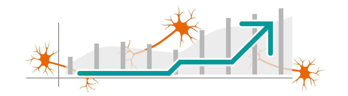
Promising therapies
- Several ground-breaking immunotherapy treatments have emerged that work to slow down cognitive decline in the early stages of AD
- These disease-modifying therapies (DMTs) are providing patients with hope for improved outcomes
A deep dive with ...
Tackling the rise of Alzheimer's disease with new therapies

Alzheimer’s Day 2026
The inaugural Alzheimer’s Day at ECR 2026 examined how imaging is transforming in the age of disease modifying therapies. The day brought together radiologists and neurologists in sessions ranging from the role of imaging to support in the diagnosis of the disease, the promise of new biomarkers to slow cognitive decline in the early stages of the disease, and the question of which patient groups are likely to be covered for these new treatment options by their healthcare systems.
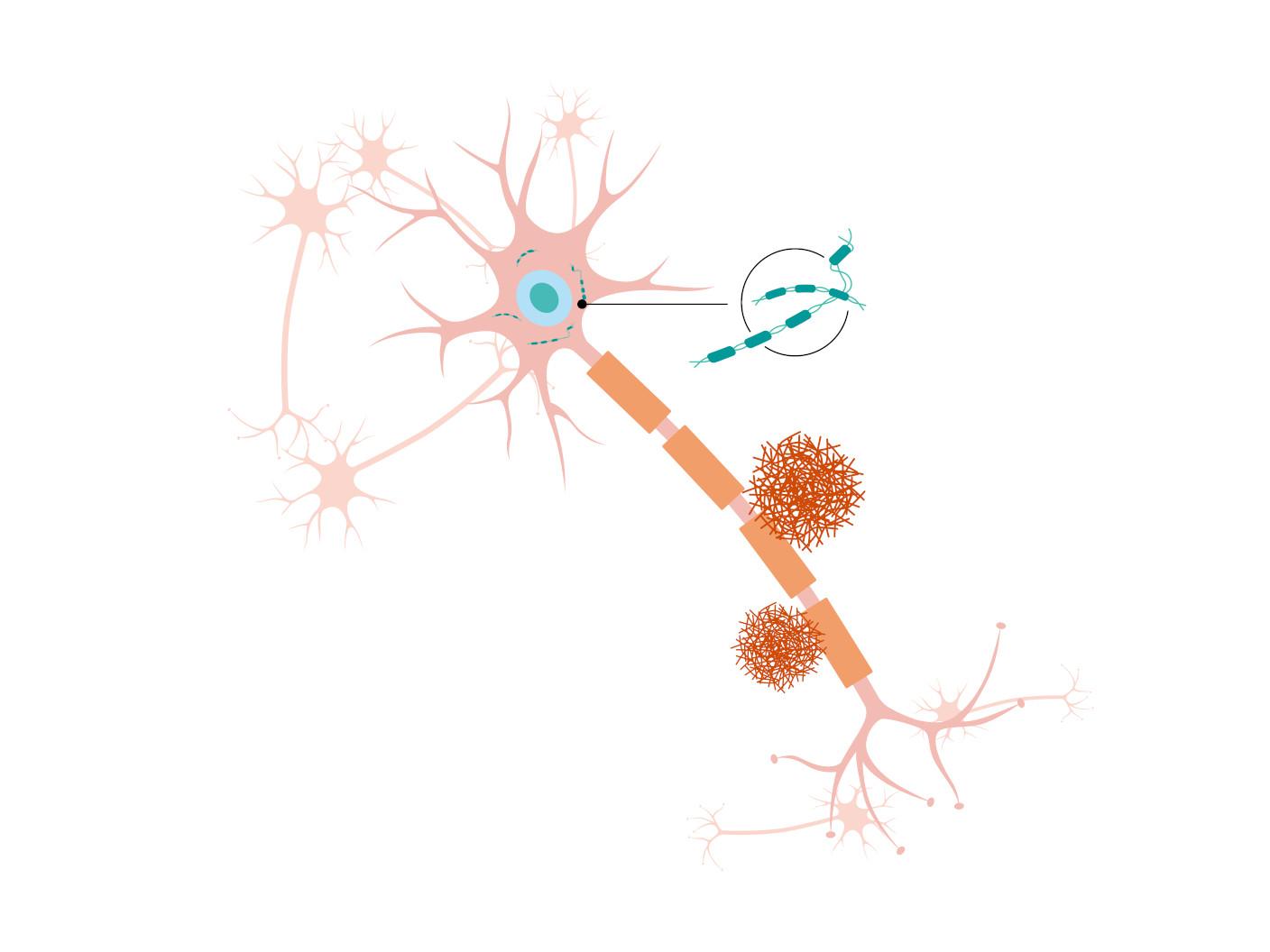
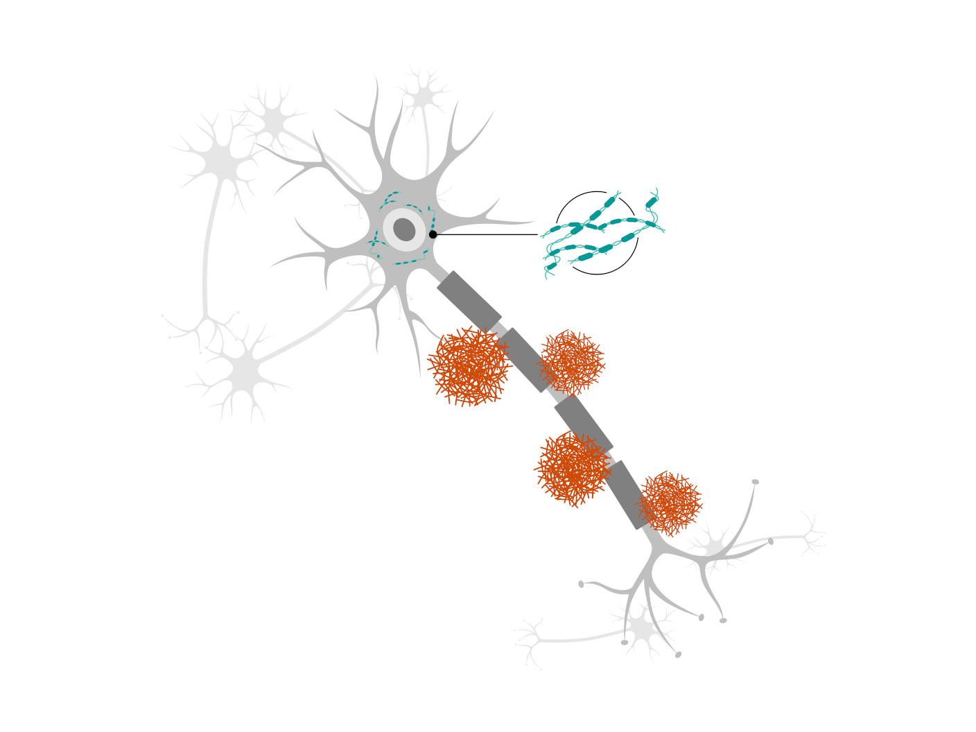
Pathology
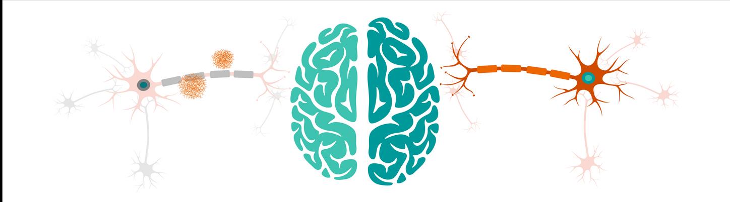
Abnormal deposits of proteins
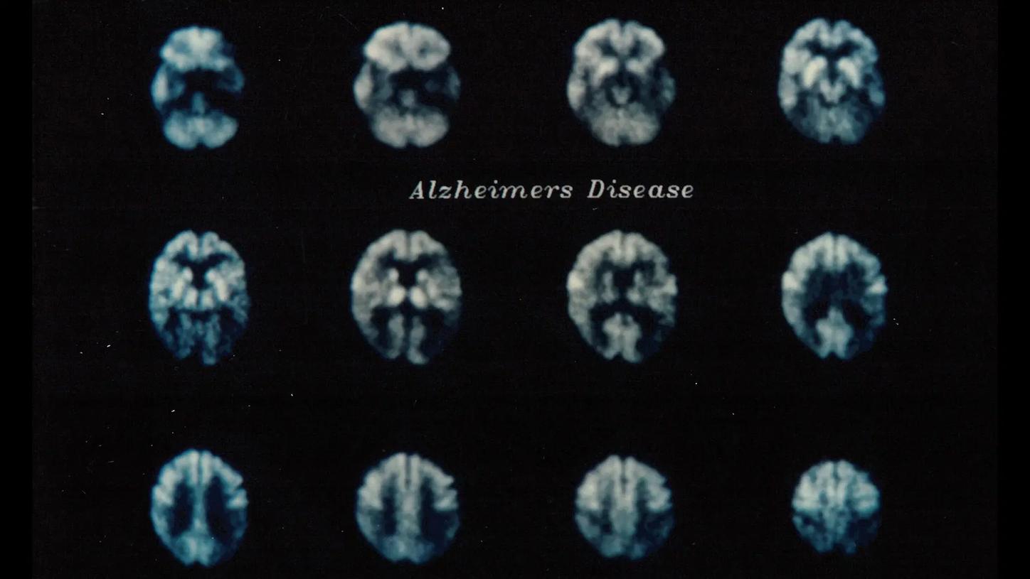
Understanding the complex brain changes behind Alzheimer's disease is essential to paving the way for new therapies. The exact mechanisms of how and why various forms of dementia develop are still being investigated. In AD, changes in the brain may begin a decade or more before cognitive decline is noticed.
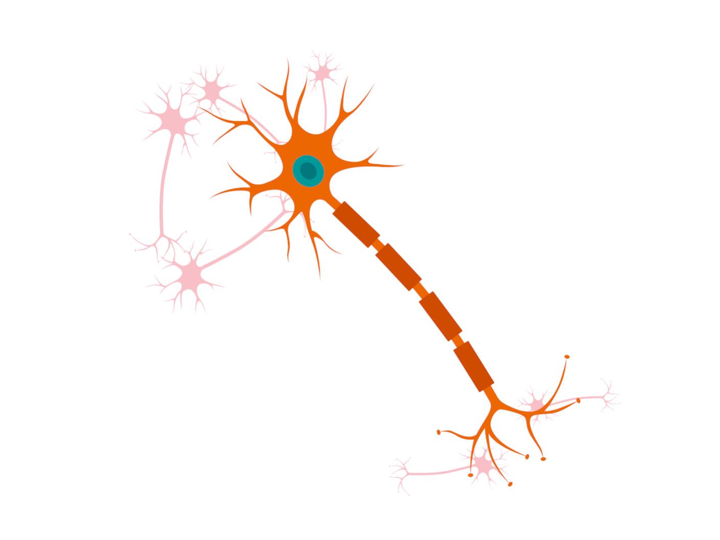
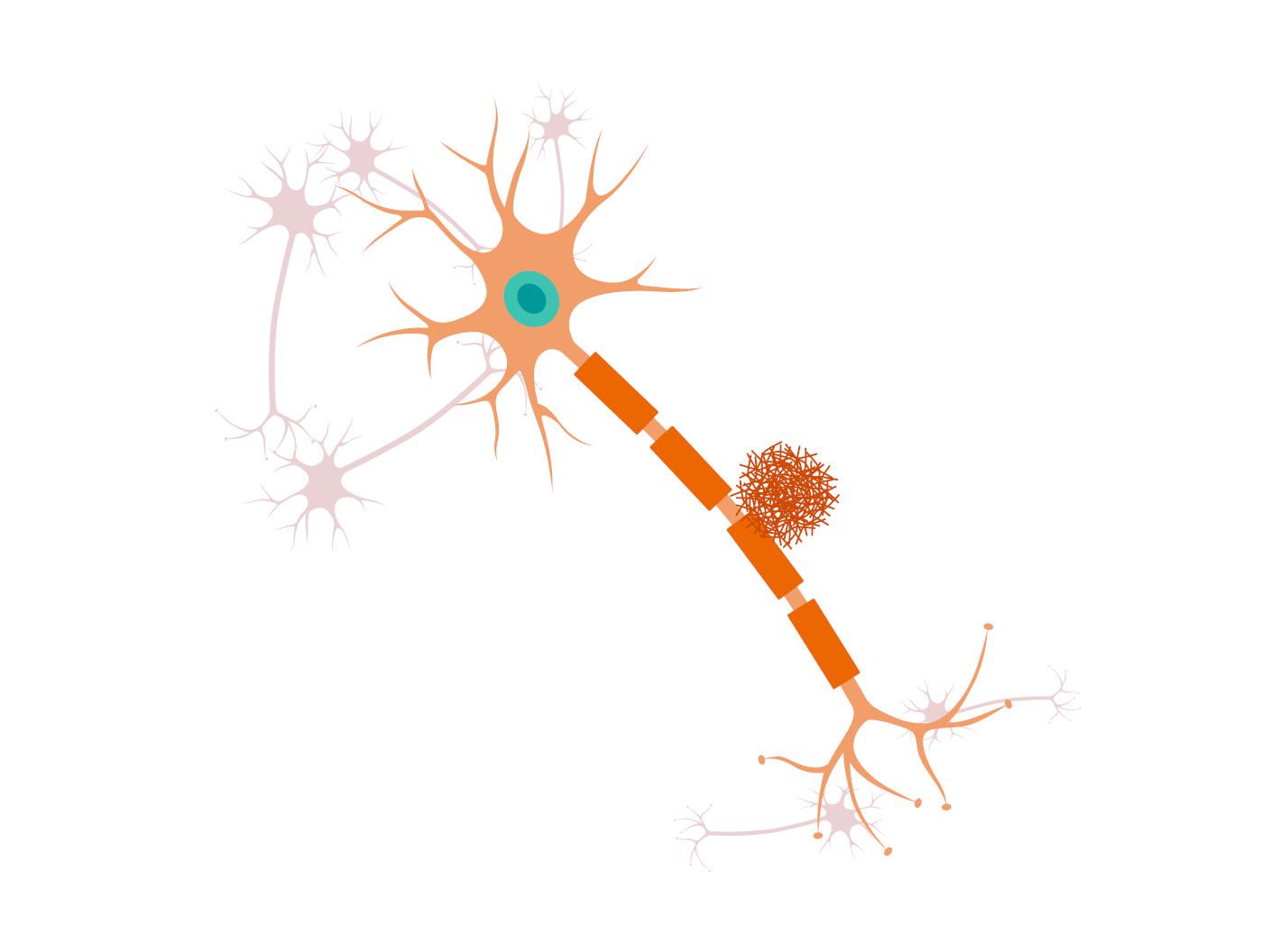
Healthy neuron
A healthy neuron without any signs of neurodegeneration
Evaluation

Diagnosis
The various forms of dementia are difficult to differentiate diagnostically. For a reliable diagnosis of Alzheimer's disease, it is necessary to:
- Assess the patient's overall health history
- Conduct cognitive testing, e.g. via the MoCa test
- Perform routine blood tests to rule out other potentially reversible causes of cognitive decline
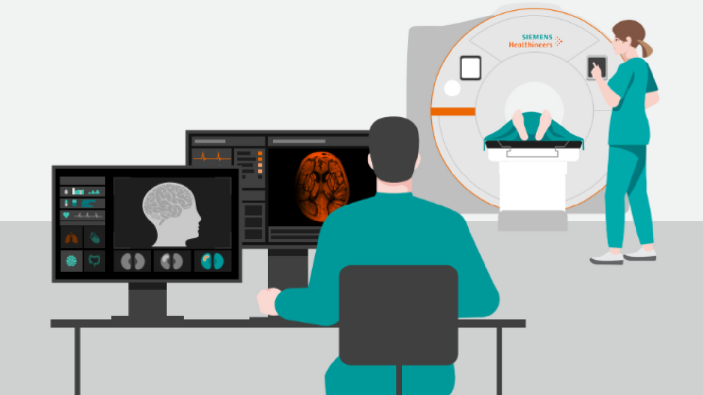
- Perform magnetic resonance imaging (MRI) to visualize structural changes in the brain (i.e. in patients with Alzheimer's disease: mesial temporal lobe and/or temporoparietal cortical atrophy)
- Perform positron emission tomography (PET) using radiopharmaceuticals to visualize amyloid and/or tau deposits, and to rule out other potentially reversible causes of cognitive decline
- Perform a lumbar puncture to analyze cerebrospinal fluid (CSF) for amyloid and tau molecules
Alzheimer's disease patient poster
Download and print our Alzheimer's disease patient poster - a clear, accessible resource designed to support awareness and understanding of Alzheimer's disease symptoms, risk factors, underlying causes, and current diagnostic and treatment options.
Treatment
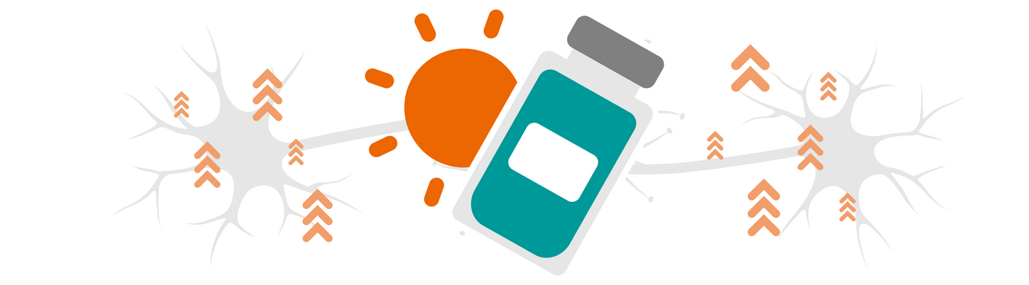
Understanding DMTs
The battle against AD has long been one of the most challenging and heart-wrenching. However, new hope has emerged—novel therapies. These DMTs4 work to slow progression of this devastating disease. Treatment is focused on patients with mild cognitive decline or mild dementia due to AD.
How do DMTs work?
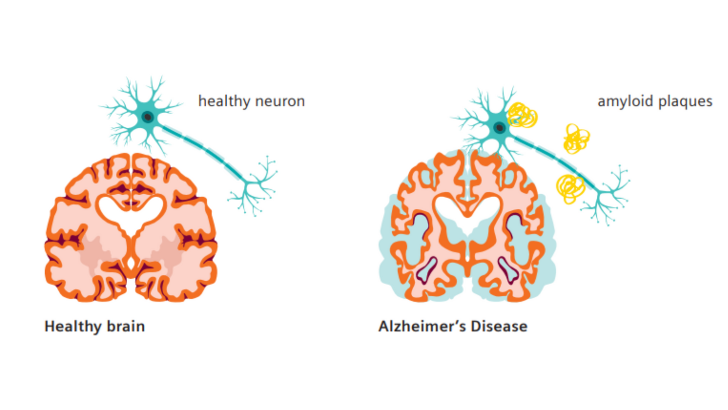
DMTs4 target the pathology of AD by reducing the beta-amyloid plaques that typically accumulate in the brains of patients with AD. Clinical studies have demonstrated that removing beta-amyloid from the brain reduces cognitive and functional decline in patients living with early AD.
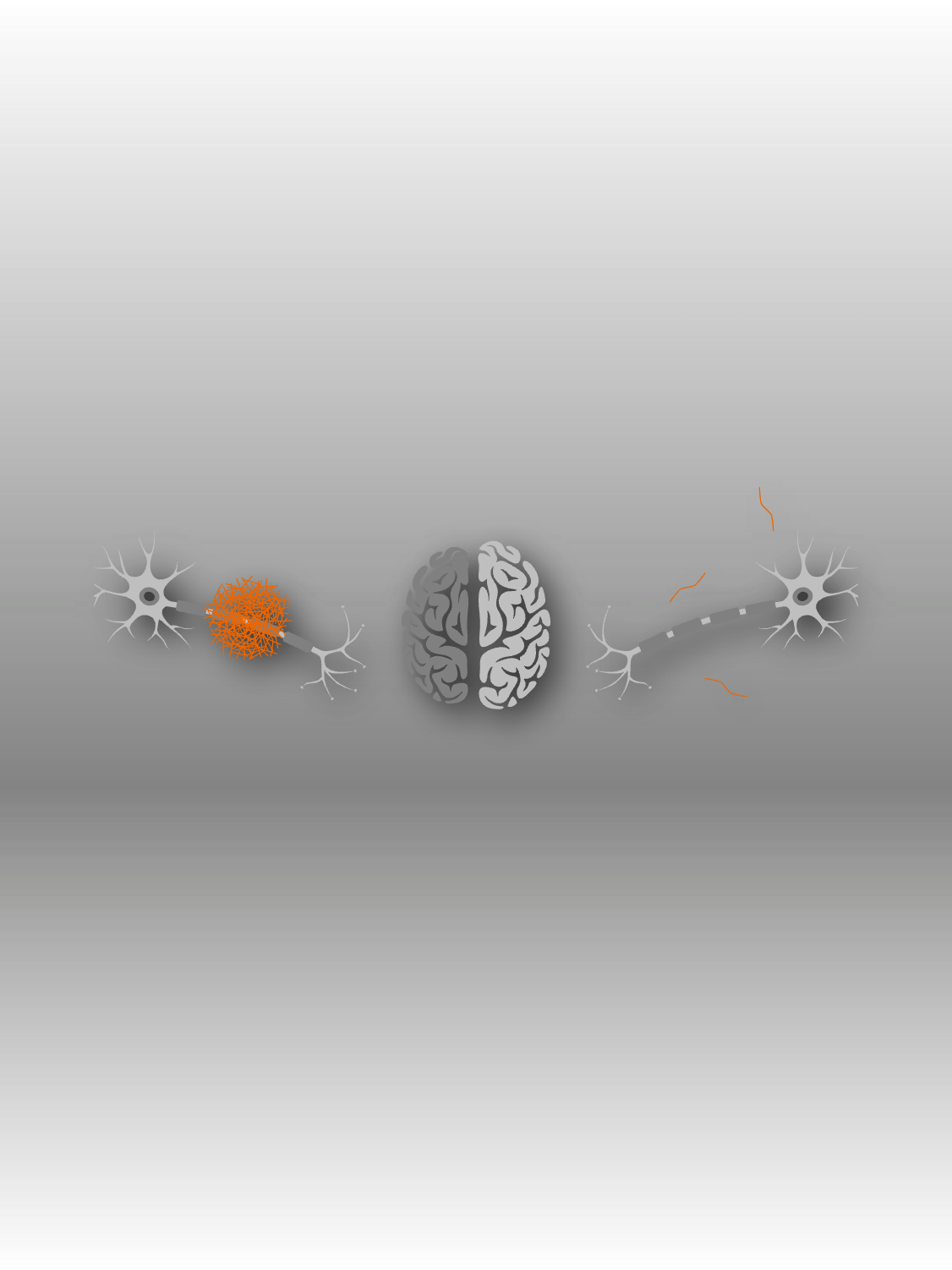
Beta-amyloid protein accumulates around brain neurons
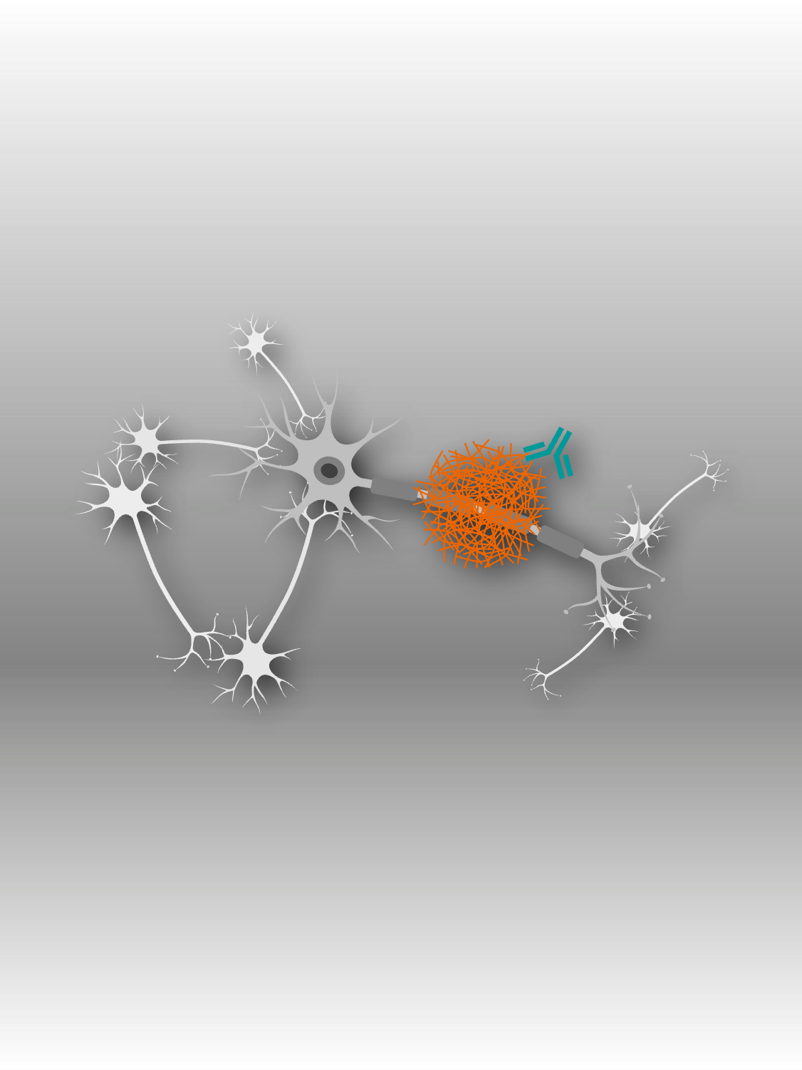
DMT antibody binds to beta-amyloid protein
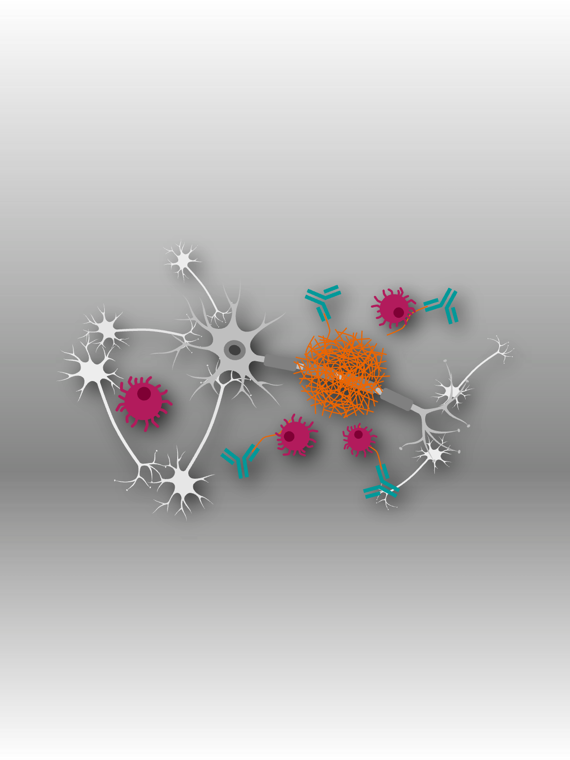
DMT antibody attracts immune cells to break down the protein
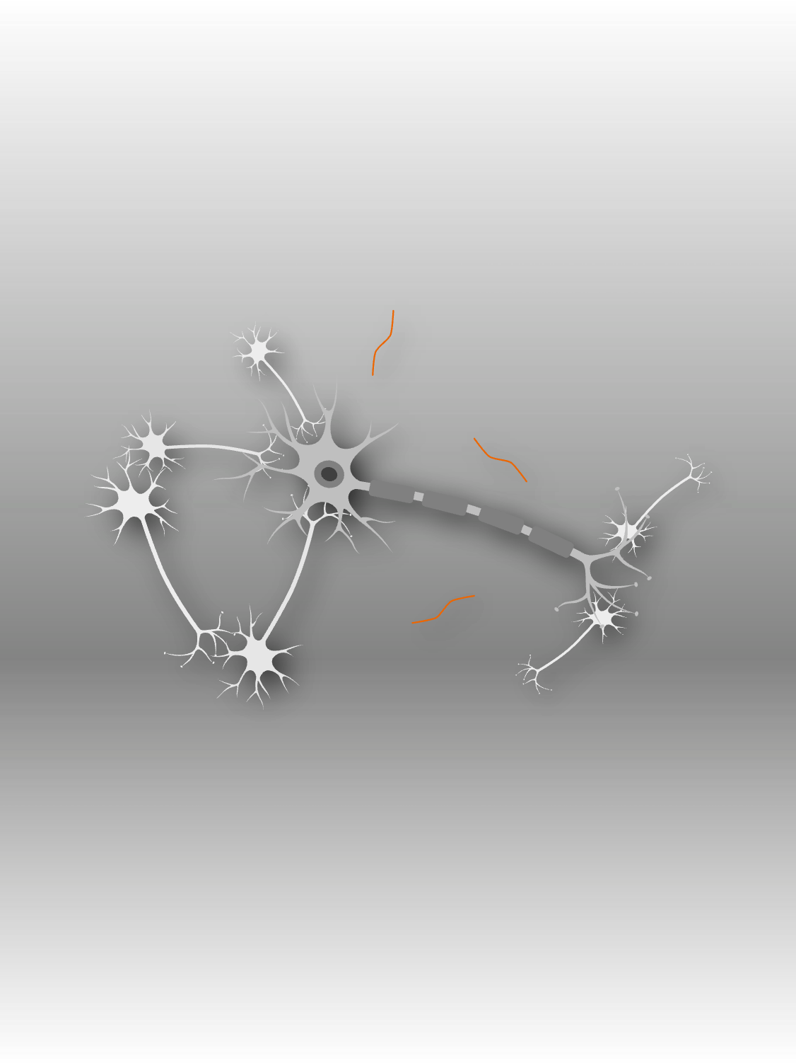
Less beta-amyloid protein around the neuron

Alzheimer’s Care Pathway
Follow Rose on her journey and discover how our digital and AI-powered solutions support scanning patients and reading images, empowering you to deliver high-quality, patient-centered care, efficiently.
Disease modifying therapies for Alzheimer's and their impact

Navigating the care pathway
These new DMTs4, while promising, require careful consideration to identify patients early enough in the disease progression:
- To enter therapy, the presence of beta-amyloid pathology must be confirmed by either cerebrospinal fluid lab diagnostics or beta-amyloid PET imaging
- MRI monitoring is necessary during treatment to monitor for adverse side effects or amyloid-related imaging abnormalities (ARIA)
Imaging is set to play a pivotal role in both early detection and the long-term management of Alzheimer's disease, providing clinicians with powerful tools to improve patient outcomes.
A clinician's guide to detecting and diagnosing ARIA in Alzheimer's disease patients
Clinicians play a critical role in precisely detecting and diagnosing ARIA to ensure patient safety and support the effective delivery of treatment.
“I have lost myself, so to speak”
Alzheimer’s: The history of the disease of forgetfulness
“As the first obvious sign of her illness, a 51-year-old woman began to exhibit jealousy toward her husband. A rapid deterioration in her memory was soon apparent, and she could no longer find her way around at home. She would move things back and forth and hide them. Sometimes she believed someone was out to kill her and began to scream loudly…”
Your partner in AD care
Explore our holistic product and services portfolio along every step of the Alzheimer's care pathway.