
syngo.via RT Image SuiteBoosting efficiency
Radiation therapy is a dynamic and fast-changing field. The number of patients receiving this type of therapy is continually rising. Software applications have a key role to play here and can support efficient workflows and deliver precision for advanced therapies, they will drive clinical excellence in RT today and into the future.
syngo.via RT Image Suite was developed for radiation oncology professionals. Designed as a user-friendly work aid to make simulation, image assessment, and contouring easier and better integrated. It simplifies and standardizes daily tasks, and gives you the capabilities you need to go beyond the current standard.
Ease what you do. Seize new opportunities.
Features & Benefits
Ease what you do
Simulation, data preparation or contouring – routine tasks can suffer from workflows across many systems, manual work or unintuitive tools. Work more efficiently and comfortably with an efficient, flexible, and well integrated tool.


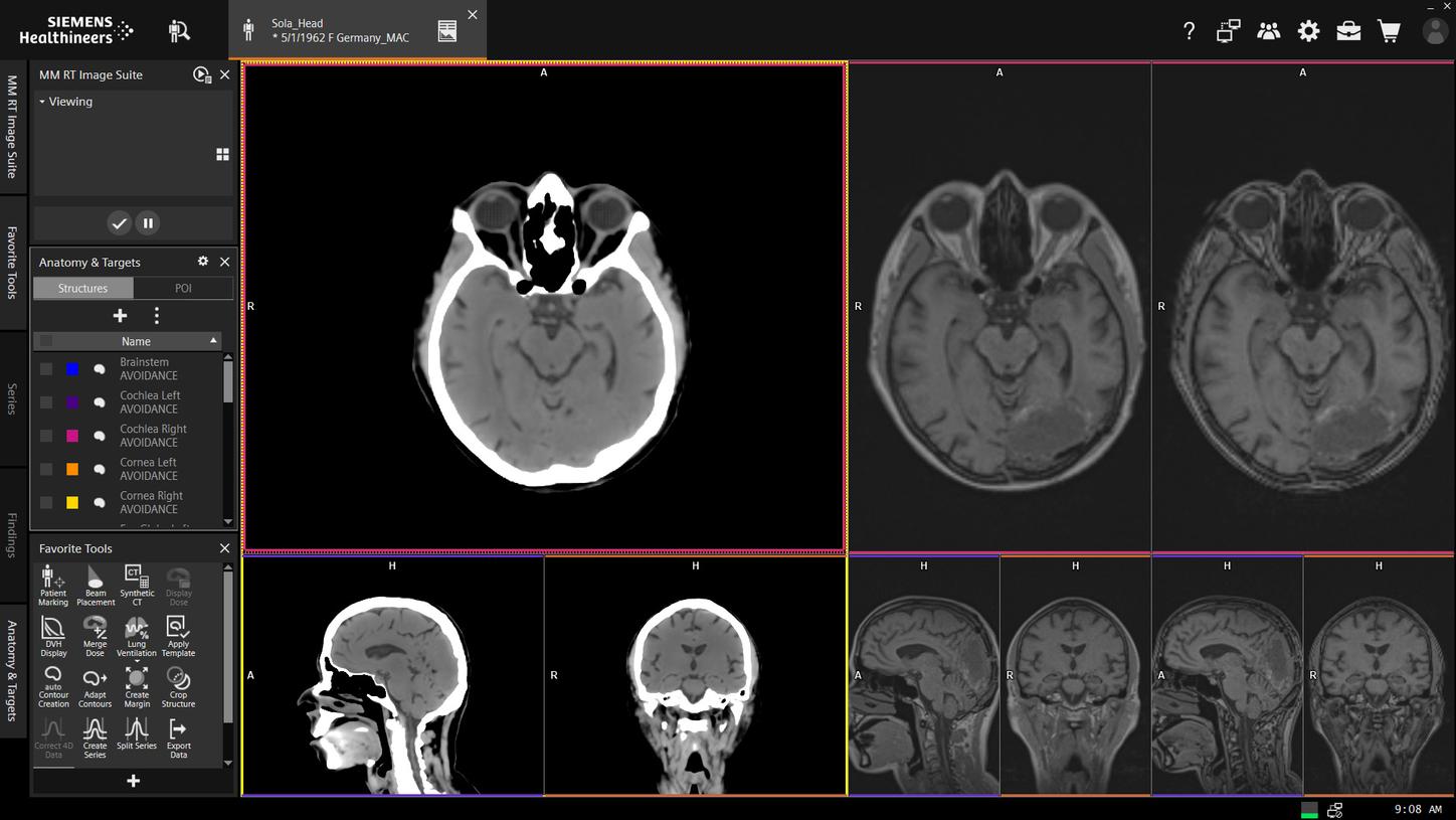
Simplify your clinical workflow with contouring tools
- Deep learning-based OAR contouring automatically provides results to your treatment planning system for CT data
- Smart contouring tools for parallel contouring on multiple images enabling "CT-free" contouring on MR or PET images
- Use multimodality images more confidently with Deformable Registration1 and Contouring Propagation1


Seize new opportunities
From introducing new treatment techniques to accurately contouring and planning with state of the art imaging or implementing inter-disciplinary cancer care – lacking tool support can make life hard for radiation oncology professionals.
Seize new opportunities and take your practice beyond today's standard with syngo.via RT Image Suite.

Courtesy of Universitätsklinikum Erlangen, Strahlenklinik, Germany
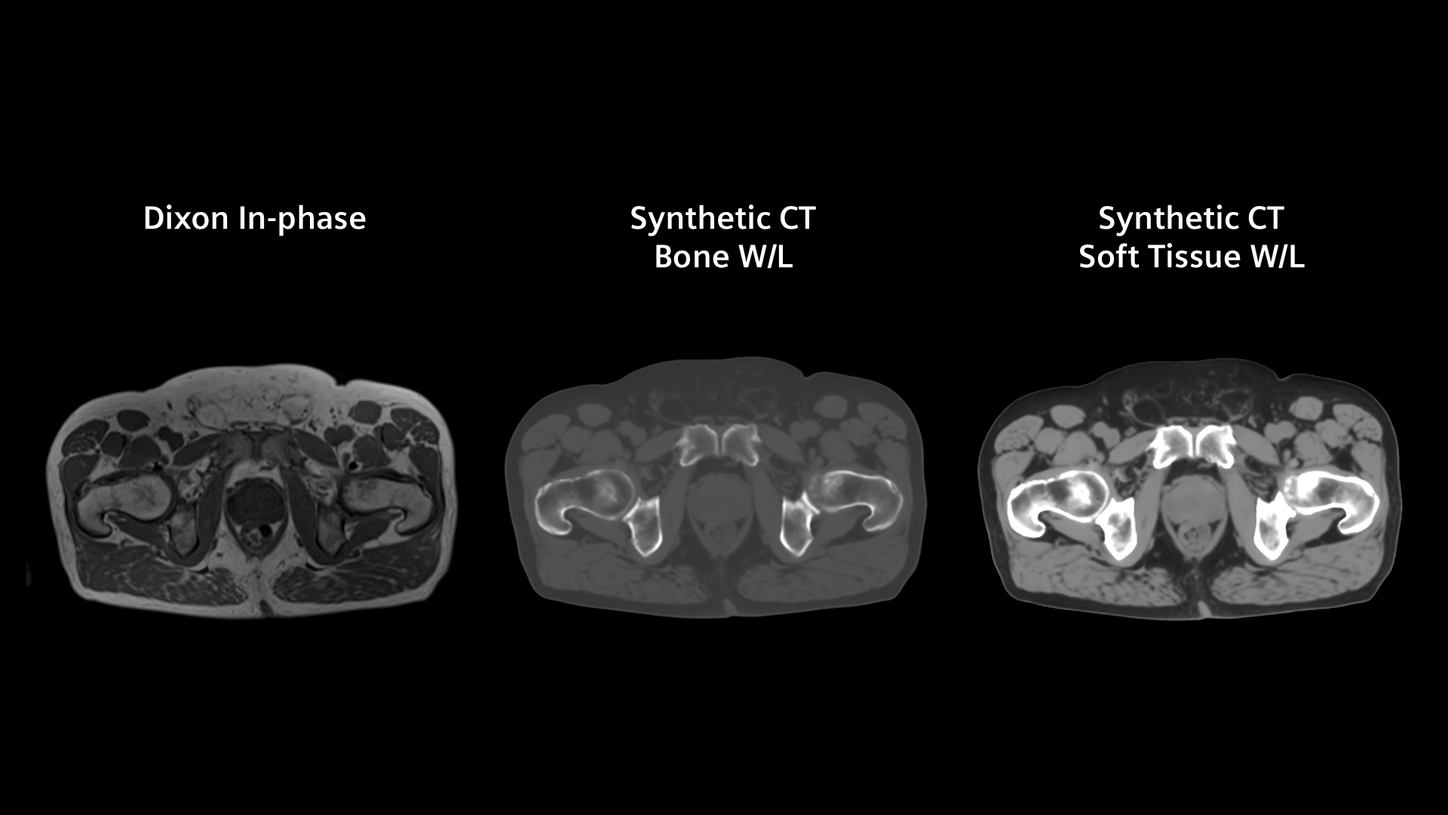
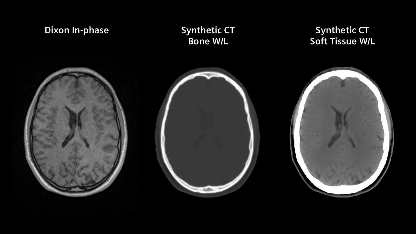
MR-only workflow with MR-based Synthetic CT6
- Use a straightforward MR-only workflow with MR-based Synthetic CT for brain and pelvic cancer patients
- Eliminate the problem of registration errors between CT and MR in radiation therapy
- Easy assessment of the alignment between the Synthetic CT and MR images by using the checkerboard


Deep-learning based autocontouring with machine learning-trained algorithms for increased efficiency and consistency in radiation therapy planning
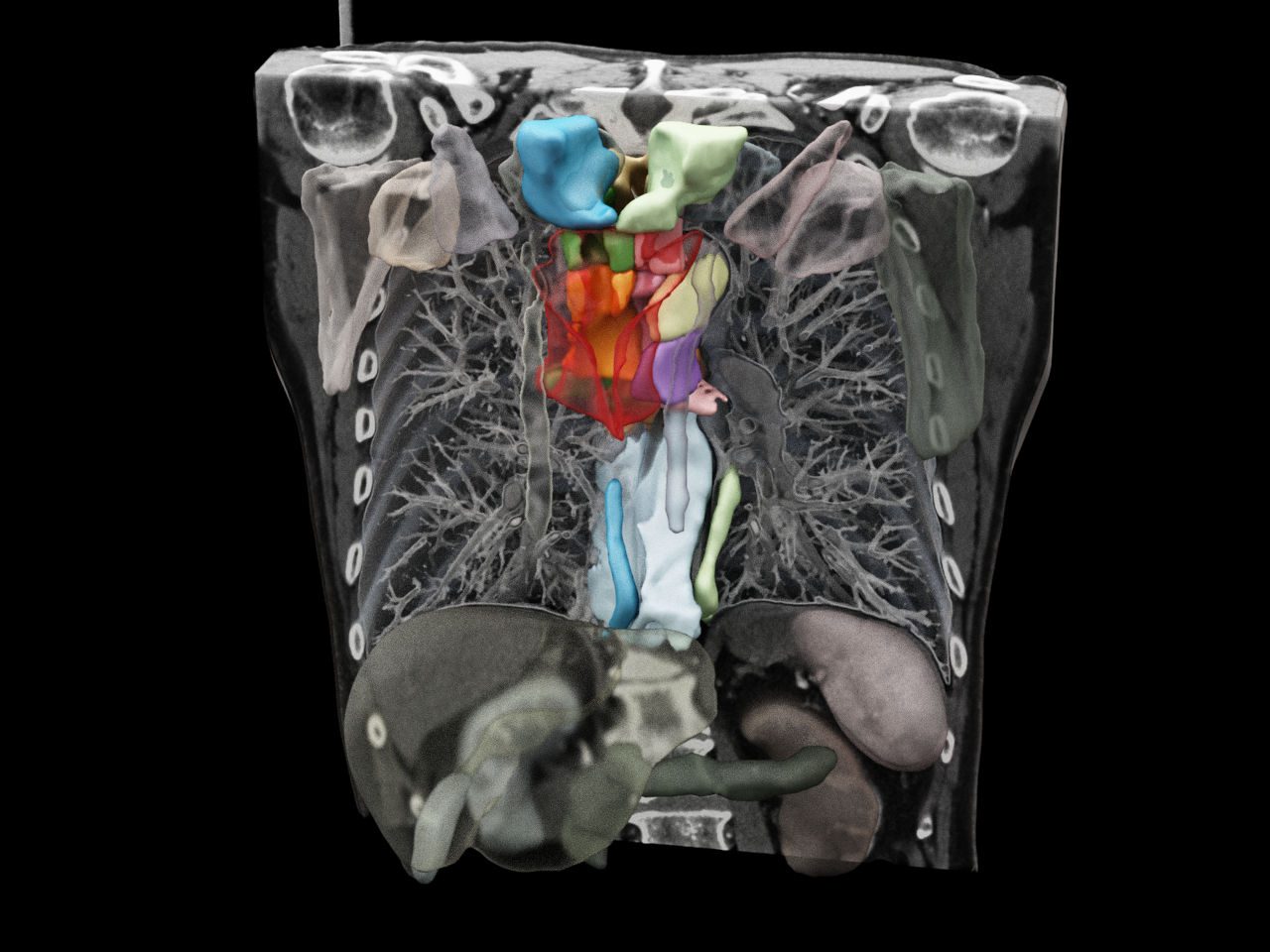
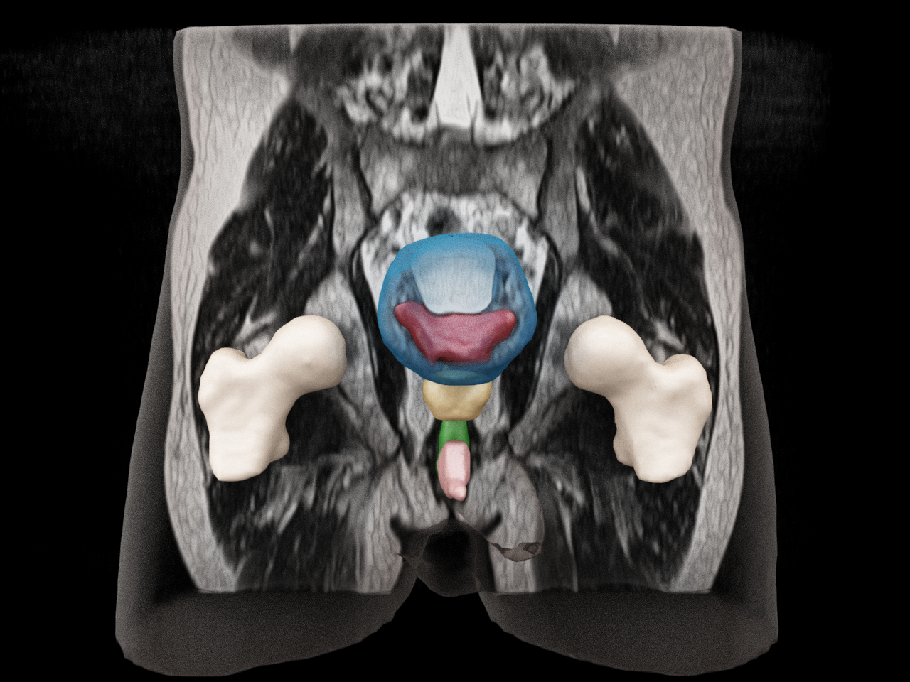
syngo.via RT Image Suite supports all relevant body regions for OAR contouring for CT data
- Brain and Head & Neck
- Thorax & Breast
- Abdomen
- Pelvic Area
- Lymphnode regions for Head & Neck, Thorax, Breast, and pelvic area
Additionally, it´s possible to customize the contouring process for specific organs (e.g. breast or brainstem) by choosing from two different contouring guidelines (e.g. ESTRO or DAHANCA)
Courtesy of Diagnostikum Graz, Graz, Austria
Autocontouring results are generated by Siemens Healthineers.
The displayed renderings are created with software which is not commercially available.
syngo.via RT Image Suite supports brain and pelvis OAR contouring and brain metastases contouring for MR data
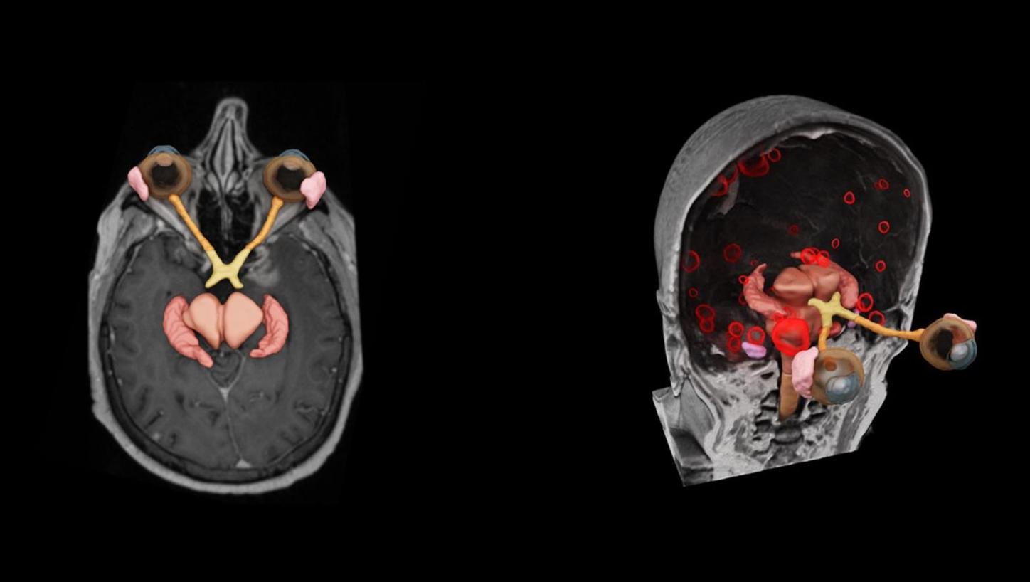
Courtesy of Universitätsklinikum Erlangen, Germany
Autocontouring results are generated by Siemens Healthineers.
The displayed renderings are created with software which is not commercially available.
MR-based Synthetic CT6
AI-powered algorithm for MR-only workflow in pelvis and brain

Challenges of combined MRI and CT workflow
- Accurate image registration
- Patient scheduling
- Resource utilization & reimbursement
MR-based Synthetic CT results for pelvis and brain
Clinical Use
Blinded evaluation of autocontouring at Universitätsklinikum Erlangen


Study details:
Clinical evaluation of clinical images from 50 patients with a total number of 2,040 organs-at-risk structures. Each resulting OAR was then evaluated by 3 physicians in a blinded study (manually vs. auto-contoured clinical images).8,11

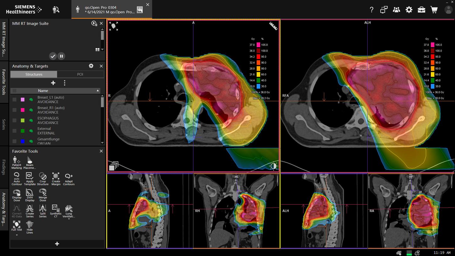
Dose distribution on MR-based Synthetic CT and CT with corresponding DVHs
- <1% mean dose difference in PTV, GTV, evaluated OAR12
- <0.5% and 1.4% mean dose difference for the brainstem and chiasma respectively12

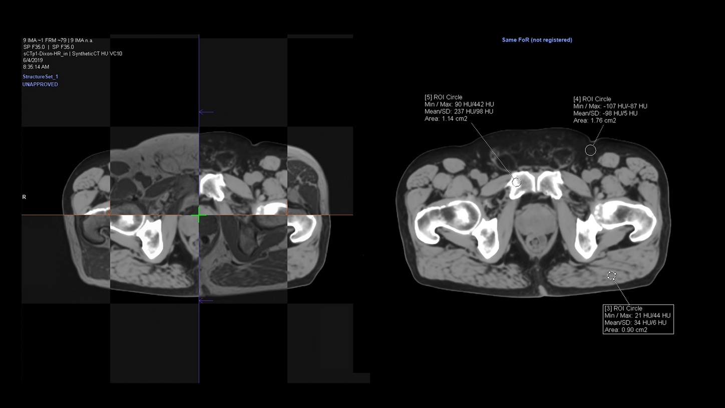
An internal validation of geometric fidelity and HU unit accuracy
The geometric fidelity test was passed with an average symmetric surface distance (ASSD) <1mm for brain and pelvis. For HU accuracy, line profiles from CT and MR-based Synthetic CT of the same patient were compared as shown in the image.12
Testimonials
“Current state of the art enables OAR autosegmentations that are on par with human experts.”11

Universitätsklinikum Erlangen, Strahlenklinik, Germany


Reduce contouring time in complex cases?
Consistent, high-quality contoured images are key for precise radiation therapy planning. The automation of organs-at-risk contouring could help increase consistency while achieving better efficiency. Gain first-hand user experience from the Department of Radiation Oncology at Universitätsklinikum Erlangen.
Siemens Healthineers and AI
Find out how Siemens Healthineers is transforming big data into precision medicine.

Did this information help you?
The products/features (mentioned herein) are not commercially available in all countries. Their future availability cannot be guaranteed.
Optional. Deformable Registration license recommended.
Interactive Spectral Imaging support will only work with a CT Dual Energy license.
Optional. MR-based Synthetic CT is an optional feature available in syngo.via RT Image Suite starting from software version VB60.
Ventilation results produced by syngo.via RT Image Suite should not be used as the sole diagnostic tool.
The case evaluation was conducted with Organs RT on syngo.via RT Image Suite.
The depicted organs are based on the selected organ template.