The Nîmes University Hospital leads the way in France with its deployment of high-end CT scanners in emergency rooms and groundbreaking research into low-dose, high-quality imaging. A visit on location revealed that the radiology unit’s spirit of innovation and success in improving patient outcomes are grounded in efficient management and dedicated teamwork.
Photos: Matthieu Colin
Download your PDF here
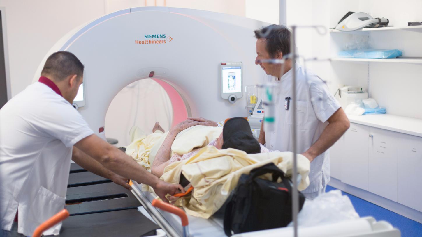
Don’t be fooled by the apparent normalcy of the radiology and imaging rooms at the Nîmes University Hospital (CHU Nîmes) in southern France. With its emergency rooms next door to the radiology unit, CHU Nîmes is quietly breaking barriers by using high-end imaging equipment on patients in need of urgent attention. That means better diagnosis with lower dosages of radiation – and improved patient outcomes.
High-end machines such as the Dual Source CT scanner SOMATOM Force by Siemens Healthineers are increasingly deployed in emergency rooms in some parts of the world. Not so in France. “It’s far from common to have such scanners in emergency rooms,” said Joël Greffier, Medical Physicist. “I think we are the among the first in France and maybe in Europe, too.”

Perennially atop the World Health Organization’s rankings for healthcare, France’s mostly publicly funded system keeps tight tabs on big ticket purchases such as high-end CT scanners. “I work in a public hospital,” said Jean-Paul Beregi, head of the radiology and medical imaging department and professor of radiology and imaging at CHU Nîmes. “The purchase must always be justified to the hospital community and beyond.”
With intensive use, the hospital’s return-on-investment begins to look pretty good. “When you buy this equipment, it is expensive. But, overall, it is cushioned by the number of patients,” said Beregi. “The cost is pretty well amortized by the current system of reimbursement in France.” In addition, CHU Nîmes serves as a regional hub – about two-thirds of CT scans are performed on patients referred from nearby partner hospitals.

Multiple missions to fulfil
Beregi began to instill a new mindset when he took over as head of radiology at CHU Nîmes in 2010. He drafted a research agenda that focused on the intersection between image quality and radiation dosage. “We made a strategic choice to be competitive internationally,” he said. Since CHU Nîmes is a public hospital, it also made sense to work on imaging applications for a broad range of specialties: cardiovascular, virtual colonoscopy, gynecology, neuroradiology, etc.
A year into his tenure, Beregi needed to replace a scanner that could be used for emergencies. So he took the chance not only to install a new system but thereby launching a cooperative relationship that included research into how to reduce radiation dosage. “Several studies have been published on how to use interactive constructs, either to improve image quality or to lower the dosage while maintaining image quality,” he said.

Testing ultra-low-dose protocols
A novel but fortuitous twist in the research plot came 2012 with a request to delve into the emerging field of virtopsy, the use of scanning and imaging technology to perform autopsies.Like everyone, CHU Nîmes had previously been using “phantoms” – defined as “hard, soft and digital materials that mimic the responses of human tissues under specific conditions,” according to the National Institute of Standards and Technology (NIST) of the U.S. Department of Commerce. The NIST specifies that these “materials include plastics, salt solutions, silicones, epoxy, polyurethane foams, carbon powder, water, disposable diapers and radioactive substances.”
For research purposes, however, none is better than a human body. Thus, virtopsy research gave CHU Nîmes carte blanche “to analyze the bodies and to look inside with the help of the scanner,” Beregi said. “That allowed us to discover and test extremely low dose protocols, which we now call ‘Ultra-low-dose’ because the dose delivered is more or less comparable to that of an X-ray.”

Better teamwork for better workflows
Beregi is also recognized as a leader in the use of advanced technologies in radiology. “Artificial Intelligence (AI) is a very broad term. And a fashionable one,” he said. “I started working with AI 15 to 20 years ago.” Machine learning and software have made significant contributions to radiology over that period, notably in terms of image quality and analysis but also in workflow management. “Not only have the number of exams increased, but also the number of images,” he said. “Yet, while the workload has grown significantly, the number of radiologists has not – so AI is of help in this regard. But if there is to be a place for AI in the future of medicine, it has to yield a guaranteed benefit for the patient. As of today, the radiologist is still the guarantor for the correct usage, the correct exam and choosing the correct software for the challenge at hand.”
Beregi hovers over the quality and quantity of diagnostic tests. The radiology and emergency departments perform about 750 examinations a day at 18 workstations, counting all modalities. Beregi holds four meetings a month to review performance indicators, including patient satisfaction surveys. “Because there are so many emergencies, there must be room in the system for flexibility,” he said. Using economic and patient data, his team looks closely at fleet- and patient management and workflow.
Beregi applies the most current organizational techniques and encourages teamwork – a somewhat novel approach in a country known for top-down management and rigid job descriptions. “We are constantly changing and adapting,” he said. “There is no standard organization that applies to my hospital or service.” He added that ‘team’ is the magic word. “If we do not work as a team, we do not do a good job. We might be good ourselves, but the other people won’t be engaged.” “Not everywhere do you find opportunities to do research and work in teams,” Greffier said. He finds it “interesting” to work on different clinical challenges and varied research topics. “We are very lucky,” he noted.
About the Author
A former correspondent in South America for The Financial Times and Business Week, Bill Hinchberger is a Paris-based freelance writer. He has contributed to publications such as The Lancet and Science, and reported for the Medical Education Network Canada.