Breast MRIHigh sensitivity and specificity to support cancer imaging

Breast MRI

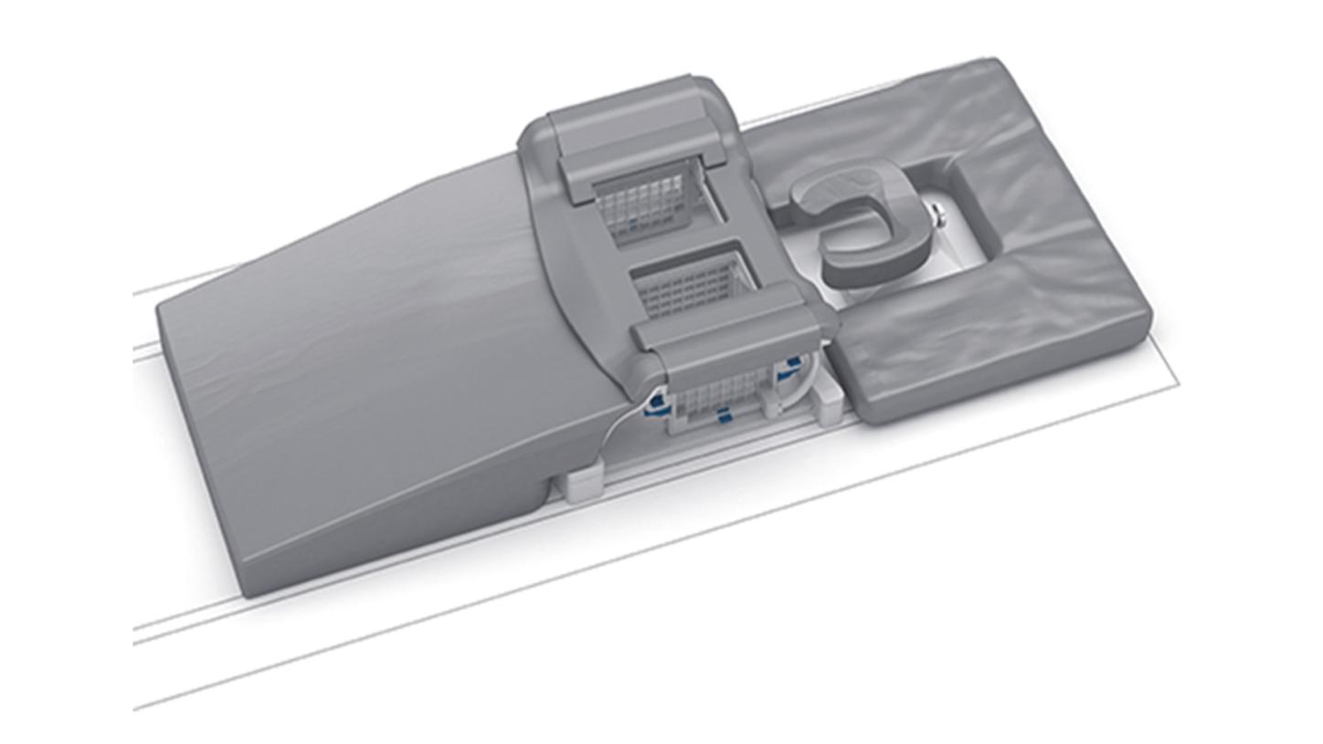
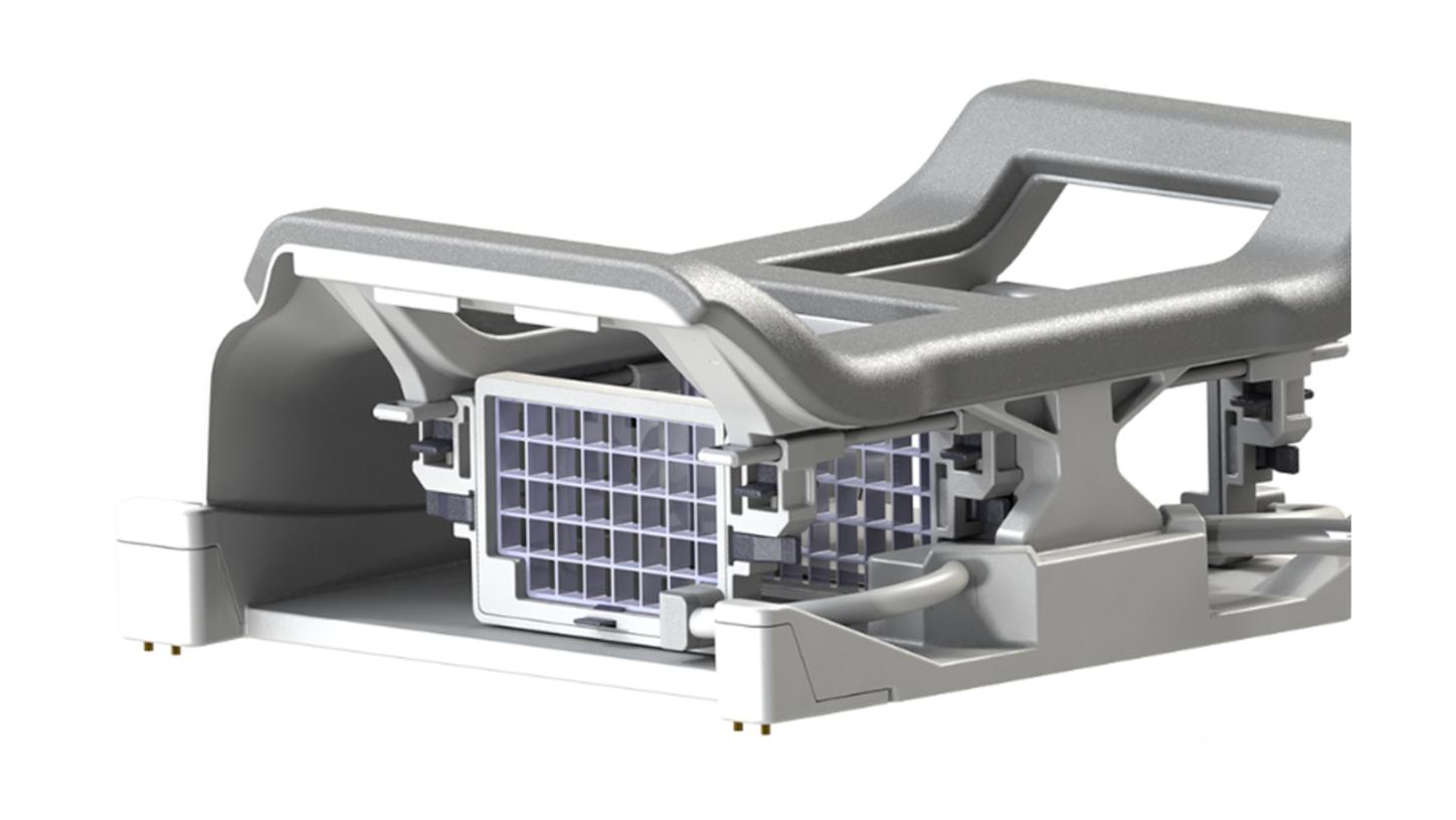
Breast cancer is the most common cancer in women. On a global scale, every tenth woman is affected, and numbers continue to rise. MR imaging can help provide early and reliable diagnosis especially in difficult cases. It can support accurate preoperative staging and differential diagnosis, resulting in faster and more cost-effective patient management.

Recent advancements such as abbreviated MRI protocols and novel techniques for functional tissue classification are designed to further the reach and diagnostic capabilities of breast MRI in clinical practice.

Applications

We provide high-resolution protocols for morphological and dynamic contrast-enhanced imaging of the breast at 1.5T and 3T. Typical challenges in the breast, such as artifacts caused by motion or susceptibility are adressed with state-of-the art hardware as well as software solutions. We offer several applications for reliable routine breast imaging and click-n-go protocols for consistent results in breast imaging, even in patients with implants. Advanced applications for functional tissue assessment, such as DWI or spectroscopy can support therapy monitoring and breast tissue visualization due to increased spatial resolution.


VIEWS: Volume imaging with enhanced water signal

VIEWS: Volume imaging with enhanced water signal

VIEWS provides isotropic 3D imaging protocols with high temporal and spatial resolution as well as excellent fat suppression. You can acquire images bilaterally or unilaterally with excellent coverage of the axillae and retrosternal regions.

TWIST: Time-resolved angiography with interleaved stochastic trajectories

TWIST: Time-resolved angiography with interleaved stochastic trajectories

TWIST is an advanced application for time-resolved, dynamic 3D imaging, based on a k-space sampling algorithm. Signal-enhanced data can be used to evaluate potential tumor vascularization. TWIST provides submillimeter spatial and high temporal resolution as the basis for high-quality dynamic datasets that can be evaluated in a 3D or 4D format.


RESOLVE: Readout segmentation of long variable echo-trains

RESOLVE: Outstanding diffusion-weighted imaging (DWI)

RESOLVE reduces blurring and susceptibility to artifacts. It is largely free of distortions and delivers sharp images at higher spatial resolution. RESOLVE is especially attractive for the evaluation of smaller lesions in a wide range of examinations.


REVEAL diffusion-weighted imaging
Different b-values and ADC map

REVEAL: Diffusion-weighted imaging

MR breast spectroscopy

GRACE: Generalized breast spectroscopy exam

Imaging without BLADE vs. with BLADE
Correction of motion artifacts

BLADE: Correction of motion artifacts

Workflow & Postprocessing

Time-consuming set-ups and post-processing can slow down all aspects of your breast workflow, including examination time. Our MAGNETOM scanners offer in-line technology that enables processing instead of post-processing, speeding up the time from image acquisition to image evaluation.


Breast Dot Engine for optimal timing

Breast Dot Engine

With the Breast Dot engine, the user is intuitively guided through the entire workflow of a breast MR examination. Certain aspects, such as positioning the imaging volume and the patient-individual detection of the contrast bolus in tissue are automated, improving consistency and quality.

Dedicated strategies are also avaliable for interventions and patients with implants. The user simply tailors the exam to each individual patient, i.e., depending on the presence of implants, and defines the scanning approach.

Breast Dot Engine for excellent implant imaging results

Your benefits:

  • Optimal timing of dynamic scans
  • Step-by-step user guidance
  • Example images and guidance text are displayed for each individual step
  • Images and text are easily configurable by the user
  • Biopsy planning software (optional) displays target coordinates of the lesion on the inroom display directly at the scanner 

Breast Biopsy Software

Our new breast biopsy software is seamlessly embedded into the scanner workflow. It provides intuitive, step-by-step biopsy planning – from target selection to visualization of the target coordinates and insertion depth using the in-room display, right next to the patient.


syngo.MR BreVis

syngo.MR BreVis

If breast imaging is part of the routine, a fast, reliable, and easy-to-use processing and workflow solution for reading and reporting is needed. Our dedicated reading and reporting software syngo.MR BreVis is a computer-aided tool for real-time breast analysis.


Your benefits:

  • Breast specific windowing presets
  • Elastic registration for patient motion correction as auto preprocessing step
  • On the fly time-course evaluation and color overlay maps
  • A statistic on lesion enhancement helps you assess therapy effectiveness
  • Ad-hoc integration of spectroscopy for lesion characterization
  • Multi-modality display
  • Reporting according to the BI-RADS standard 

Coils

We support breast imaging on all scanners of the MAGNETOM family and offer breast coils to meet different needs. Our breast coils are designed to provide high SNR for excellent diagnostic image quality or to provide greatest access to the breast for interventions.


The features:

  • 18 independent channels for exceptional iPAT capabilities
  • Only 5.5 kg weight and SlideConnect
  • Mechanically adjustable stabilization padding
  • Flexible up to DD cup size
  • Head first positioning is possible for 60 cm as well as for 70 cm
  • Feet first positioning is possible for 70 cm


Breast 18 Coil Feed First

Your benefits:

  • High-speed image acquisition
  • Excellent comfort for all patients
  • Easy handling and installation for technologist 


The features:

  • 7-ch design for imaging and biopsy with high SNR
  • LED lights provide excellent visibility for breast positioning and biopsy
  • Open, spacious design for excellent biopsy access (lateral and medial)
  • Medio-lateral and optional cranio-caudal compression
  • Both grid and post/pillar biopsy supported
  • Patient-adaptive cushions for improved comfort
  • Excellent signal from the axillary region
mri_breast-bi-7-coil_post-pillar-biopsy

Your benefits:

  • Accommodates breast volumes up to 11 litres
  • Compactness and light weight (9 kg) for easy handling
  • Easy access for exact positioning
  • Always 7 receive channels (imaging and biopsy) for uncompromised image quality 

Breast Matrix Coil for imaging

Breast Matrix Coil for imaging

4-channel BI Breast Coil for biopsy and routine imaging

4-channel BI Breast Coil for biopsy and routine imaging

2/4/8-channel Sentinelle Breast Coil for biopsy and imaging

2/4/8-channel Sentinelle Breast Coil for biopsy and imaging

2/10/16-channel Sentinelle Breast Coil for biopsy and imaging

2/10/16-channel Sentinelle Breast Coil for biopsy and imaging

Sentinelle Breast Coil for MAGNETOM ESSENZA

Sentinelle Breast Coil for MAGNETOM ESSENZA

Users

Listen to and read what our Breast MRI users have to say.


Listen to the clinical talk from the ECR Breast Care Day 2014 in Vienna, Austria by
Ritse M. Mann
Radboud University Nijmegen Medical Center
Nijmegen, Netherlands

Listen to the clinical talk from the ECR Breast Care Day 2014 in Vienna, Austria by
Dr. Clemens Kaiser
University Medical Centre Mannheim
Mannheim, Germany

Listen to the clinical talk from the ECR Breast Care Day 2014 in Vienna, Austria by
Dr. Frank Stoeblen
Gynecological Radiology
Essen, Germany

Listen to the clinical talk from the ECR Breast Care Day 2014 in Vienna, Austria by
Linda Moy
NYU Cancer Institute, Breast Imaging Center
New York, USA


“MR breast imaging is a good possibility for all patients. With MR you can see a lot more than with mammography or ultrasound. It basically shows the whole extent of the disease.”

Prof. Dr. Chantal Van Ongeval
Radiologist
University Hospitals Leuven, Belgium
Siemens International Reference Center for MR Breast Care


Have a look at the MR-guided biopsy video, showing the whole procedure from equipment preparation, patient positioning, biopsy path planning, performing the biopsy, verification & sample handling, and the clip insertion as well as the localization for surgery. The biopsy is performed by
Dr. Evelyn Wenkel
University Hospital Erlangen
Erlangen, Germany

1