
September 10, 2013 | Medical Imaging is becoming vastly more important to orthopedic and trauma surgery: a trend that changes the educational requirements in this broad medical field. Impressions from the AO Davos Courses 2012.
The first thing you notice is the noise. The cavernous conference hall is ringing with the sounds of hammering, drilling, and metal being beaten into shape. Countless screws and plates are arranged on worktables, where dozens of young men and women are at work putting them together. If you didn’t know better, you might think you were in the middle of an enormous metal factory. But of course we do know better: this is one of the many hands-on training courses for orthopedic surgeons on offer at the AO Davos Courses (AO is a German abbreviation for a study group looking at questions relating to osteosynthesis). A sophisticated two-week event, held each year in Davos, at the beginning of December. For the AO Courses 2012, 1,500 surgeons have traveled from 74 countries to the Swiss alpine city, which is perhaps best known for its role as host of the World Economic Forum.
The surgeons have traveled to expand their medical training. They are here to attend the numerous orthopedic trauma, spine, veterinary, and craniomaxillofacial training courses and lectures, which are at the heart of the AO Davos Courses event. The AO Foundation – established in 1958, and today the world’s largest medical network – is “built on a commitment to advance medical education for better patient care,” says Jaime Quintero, the AO Foundation’s current president.
AO Foundation and Siemens Healthcare: An Important Partnership
Siemens Healthcare has always maintained a special relationship with this renowned organization. As exclusive imaging partner, the company has been supporting the training and education provided by the AO Foundation since 2007. With good reason: medical imaging is vastly gaining significance to orthopedic and trauma surgery. This trend is increasingly influencing the courses on offer in Davos, as Urs Rüetschi, Director of AO Education points out: “Imaging expands the scope of the whole educational program we have to create, because surgeons need to understand ever more about this technology and how to deal with it.”
Or as AO Foundation President Jaime Quintero puts it: “Nowadays, imaging is a tool for a surgeon, just like the scalpel, a clamp, or a screwdriver. It belongs to the basic set of instruments in the operating room.”
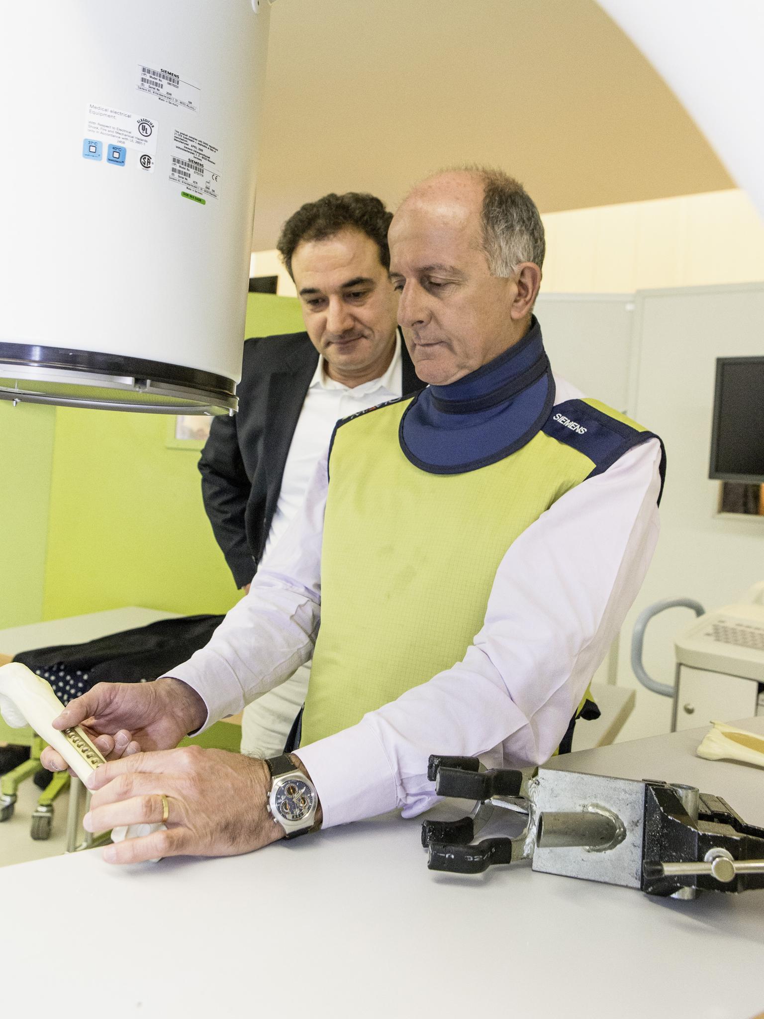
Tuesday, December 4, 12:30 in the “Aspen” room of the Davos Congress Center: Professor Martinus Richter, head of the foot and ankle surgery departments at Nuremberg and Rummelsberg hospitals in Germany, is giving a lecture on intraoperative 3D imaging in foot surgery with mobile C-arms. Introducing his topic, Richter explains that he has conducted more than 3,000 intraoperative 3D scans: the vast majority of them exclusively on feet. When asked the reason for his reliance on technology, the 43-year-old physician – who specializes in trauma surgery as well as orthopedics – replies, “Intraoperative 3-dimensional visualization provides useful information in foot and ankle trauma care that cannot be obtained from plain films or conventional C-arms.”

Surgical Challenges
This is also the conclusion that Richter and his colleagues drew from a study they conducted in 2003 and 2004, when Siemens’ ISO-C 3D system was used for intraoperative visualization in 62 cases. Nowadays, Richter uses a Siemens ARCADIS Orbic 3D in his department. He tells his audience that the main problems in foot surgery are the malpositioning of implants and incorrect reduction (repositioning of a fracture). Foot surgery is complex because the anatomy of the foot is complex: it is composed of 25 bones and contains many three-dimensional structures: “Therefore, without seeing, even a very experienced surgeon cannot be sure whether his positioning of extraosseous or intra-articular screws is correct,” he explains.
During his study, Professor Richter first used a conventional 2D X-ray to judge the reduction and implant position. Then he performed a 3D scan with the Siemens ISO-C 3D. Based on the 2D images the surgeon would have decided that the positioning is correct. But in 39 percent of cases the 3D scan revealed that it was necessary to correct the reduction and/or implant position: “However, this did not prolong the operation unnecessarily,” Richter adds. On average, operations were interrupted for 330 seconds: 120 seconds for the scan, and 210 seconds for evaluation of the images by the surgeon. “This is much less than if another surgery were needed,” he notes.

Obvious Benefits of Intraoperative 3D Imaging
In everyday practice, however, there are trauma clinics where even a control scan is not carried out following surgery. One reason for this is the reimbursement policy: the DRG system (diagnosis-related group, a system to classify hospital cases), which, for example, German and Swiss hospitals use to run their billing, does not reward the quality results that can be achieved by using intraoperative imaging: “A case is a case, whether you operate well or not,” Richter says bluntly. On the other hand, if a patient has to come back for re-operation because his or her fractured ankle has remained crooked, the DRG system does not sanction this. Instead, it regards the second surgery as a “new” case.
The other reason that intraoperative 3D imaging has not been accepted as quickly as Richter once imagined is a scientific one: there is no randomized prospective study that proves the effectiveness of the procedure. The reason why some hospitals do not conduct a study is simple, Richter explains: “In my hospital, for example, the ethics committee didn’t believe that such a study was even necessary. They thought the advantages of the device were so convincing it would have been unethical not to use it for the control group.”
Nevertheless, Richter is convinced that intraoperative 3D imaging will prevail in the longer term – providing benefits not just to foot surgery, but also to many other orthopedic fields: the spine, hands, joint fractures, “Wherever you have three-dimensional bone structures and the position really matters,” he notes. Richter even believes that in 20 years, conventional C-arms for 2D scanning will not exist anymore: “Scanning in the operating rooms will develop much as navigation systems in cars did,” he predicts. “At the beginning of the nineties, satellite navigation was rarely seen in cars, and it worked very poorly; whereas nowadays, you couldn’t imagine a modern vehicle without this excellent device.”

and Ankle Surgery Departments at Nuremberg
and Rummelsberg hospitals, Germany
A Win-Win Situation
At the AO Davos Courses 2012, participants are offered several opportunities to become acquainted with intraoperative 3D imaging: not only through lectures, but also in practical hands-on courses and experiments. For Siemens, the two-week event provides a tremendous chance to share its know-how directly with customers. Dr. Alexander Grafenberg, Siemens’ representative in Davos, is very satisfied about the company’s strong presence at the AO Courses: “In Davos, with its unique concentration of surgeons from all over the world, we have a fantastic opportunity to bring imaging into focus as one of the rising topics in surgery,” he says, noting that medical imaging used to be only of minor interest for trauma and orthopedic surgeons. In recent years, however, Grafenberg has observed a definite change in attitudes. He believes that the collaboration between Siemens and the AO Foundation is partially responsible for this trend.
AO Education Director Urs Rüetschi confirms that Siemens’ commitment has contributed substantially to the arsenal of educational material available for the training of (future) trauma and orthopedic surgeons. “In the future, we want to prove that our courses will lead to a change in practice and better performance,” he says. “This will then, of course, result in better patient outcomes.”
The AO World in Davos
From December 1 through December 14, 2012, the AO Foundation provided 21 orthopedic trauma, spine, craniomaxillofacial, and veterinarian courses to more than 1,500 surgeons from 74 countries. The surgeons were taught by 422 expert faculty members from around the world. The AO World comprises four clinical divisions: AO Trauma, AO Spine, AO Craniomaxillofacial, and AO Vet. The annual Davos Courses are the highlight of the organization’s activities, and they are highly valued by surgeons from numerous countries. Besides this major Swiss event in December, the AO Foundation is globally active throughout the year. It currently offers more than 690 courses and seminars worldwide, including those for operating theater personnel; various faculty education programs; as well as a comprehensive online and face-to-face program, aimed at improving the teaching skills of AO Faculty members.
Author’s bio
Irène Dietschi is an award-winning Swiss science and medical writer.