By Partha Ghosh, MD1, Dale L. Bailey, PhD2, Georgios Angelis, PhD2, and Paul J. Roach, MBBS, FRACP2
Data and images courtesy of Royal Northshore Hospital, Sydney, Australia
Background
Cancer during pregnancy is rare but poses major challenges regarding imaging protocols for minimizing fetal radiation exposure, especially in relation to PET/CT imaging. Although there is no consensus about the maximum radiation exposure considered safe for a fetus, the recommendation is to ensure fetal dose remains below 100 mGy.3 Because pregnant cancer patients may require multiple PET/CT studies, it is critical to optimize the PET/CT protocol to enable as low an injected dose as possible and ultra-low dose (ULD) CT for attenuation correction.
Using long axial field of view (LAFOV) PET/CT systems like Biograph Vision Quadra helps achieve high quality PET/CT acquisition with very low radioactivity due to the extraordinarily high sensitivity leading to high number of detected events. The Biograph Vision Quadra with 106 cm axial field of view and ultrafast 228 ps time of flight (TOF) performance ensures the highest detection rate capability, enabling very low radiopharmaceutical dose for optimized PET/CT imaging without increasing acquisition time.*
* Based on bench testing (e.g., improved sensitivity and ToF per NEMA NU-2:2018). Please refer to the approved PET drug prescribing information for dosing and administration instructions.
History
In routine clinical use, administered radioactivity can be decreased by ~40% - 50% while simultaneously decreasing PET acquisition times to 5 minutes for all subjects for static, delayed scans. In standard clinical practice, 150 MBq (~4 mCi) is administered to all subjects independent of patient weight or BMI. The short acquisition time gives more flexibility than a standard field of view PET system to further vary administered radioactivity and acquisition time.
Using ULD protocols, the Fludeoxy-glucose F 18 injection (¹⁸F-FDG) administered was reduced to around one quarter of the standard amount (40 MBq), while scan time increased fourfold to maintain approximately the same number of total acquired events. A 20-minute scan is similar to the duration of most clinical PET scans and is not overly burdensome for patients. The radiation dose from the ULD CT protocol was 0.2-0.3 mSv and the radiation dose from 1 mSv/50 MBq. As a result, the total radiation exposure from the ULD protocol was around 1 mSv to both mother and fetus, a level not previously thought possible.
Two clinical case examples below demonstrate ULD imaging in pregnant patients using Biograph Vision Quadra. These patients presented with breast carcinoma and lymphoma and were both approximately 18 weeks into pregnancy.
Findings: Case 1
A 34-year-old woman in the second trimester of pregnancy (16 weeks gestation) with recently diagnosed HER2 positive breast cancer was sent for 18F-FDG PET/CT study for initial staging. Clinical and ultrasound examination revealed a multifocal right breast mass with enlarged axillary lymph nodes.
The patient was injected with 56 MBq (~1.5 mCi) 18F-FDG (weight 74 kg; ~0.75 MBq/kg).* 55 minutes following radiopharmaceutical injection, a modified whole body PET/CT study was acquired from vertex of skull to mid-thighs, with the arms raised. ULD CT was initially performed for attenuation correction utilizing the Biograph Vision Quadra’s Sn filter capability with very low mAs (kVp =Sn140 mAs = 6). PET acquisition was for a single 106 cm axial FOV bed position for 15 minutes. PET was reconstructed using UltraHD PET (PSF+TOF) 4i5s with a post-reconstruction 3D Gaussian filter of FWHM = 2.0.
As shown in Figure 1, PET/CT images show multiple 18F-FDG-avid lesions within the lateral right breast, corresponding to the biopsy-proven breast carcinoma. The largest lesion just lateral to the nipple measures 1.9 cm maximum diameter with SUVmax = 12.9. Another focal lesion in the lower outer quadrant measures 1.65 cm with SUVmax = 14.3. Several smaller hypermetabolic lesions are visualized in the inner quadrant, with the largest showing SUVmax = 8.2.
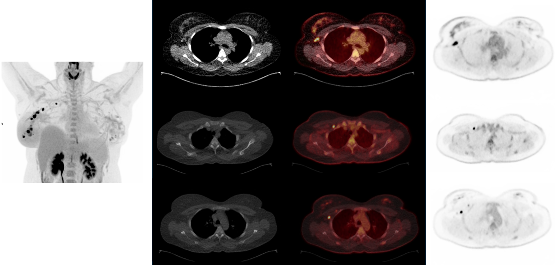
Figure 2 shows multiple hypermetabolic axillary lymph nodal metastases, with the largest measuring 18 x 9 mm with SUVmax = 14.6 at level 1. There are multiple hypermetabolic nodes located at level 2 posterior to the pectoralis minor muscle, with the largest and most 18F-FDG-avid with SUVmax = 14.3. The most superior lymph node is in the level 3 station (medial and just superior to pectoralis minor) and is immediately anterolateral to the head of the first rib with SUVmax of 10.5.
Both breasts also show physiological proliferation of parenchyma as is expected during pregnancy. No other abnormal distant foci were visualized.
The study was performed with extremely low injected dose with a 15-minute vertex-to-mid-thigh acquisition. The acquisition time was adequate for producing very high-quality images without undue patient motion despite such low injected activity.
* Based on bench testing (e.g., improved sensitivity and ToF per NEMA NU-2:2018). Please refer to the approved PET drug prescribing information for dosing and administration instructions.
Case 1
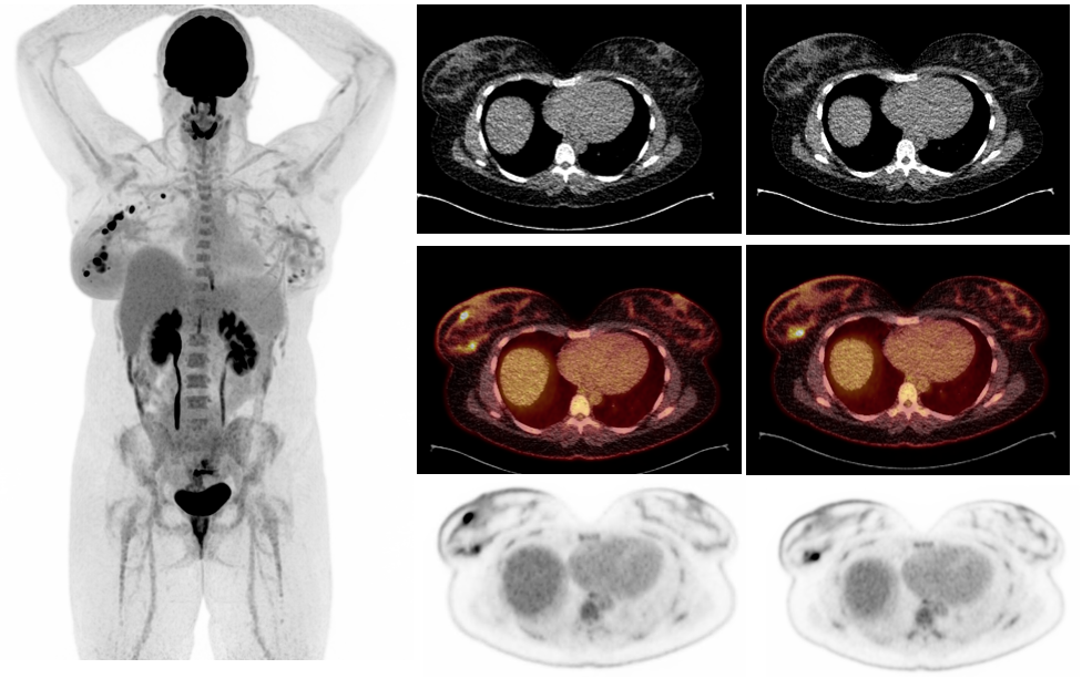
Figure 1: PET MIP and axial CT, PET, and fused PET/CT images at the primary breast tumor show multiple FDG avid masses in the right breast indicating multifocal primary breast carcinoma with multiple hypermetabolic right axillary lymph node metastases.
Findings: Case 2
A 37-year-old female patient with newly diagnosed Hodgkin’s lymphoma who was in her second trimester of pregnancy (18 weeks gestation) was referred for 18F-FDG PET/CT for initial staging.
The patient was injected with 39.1 MBq (1.0 mCi) 18F-FDG (weight 60 kg; 0.65 MBq/kg ).* 60 minutes following radiopharmaceutical injection, a whole body PET/CT study was acquired from vertex of skull to mid-thighs with the arms raised. ULD CT was initially performed for attenuation correction utilizing the Sn filter capability of the CT in Biograph Vision Quadra with very low mAs (kVp = Sn140 mAs = 6). PET acquisition was for a single 106 cm PET axial FOV bed position for 20 minutes. UltraHD PET reconstruction using 4i5s with a post-reconstruction 3D Gaussian filter of FWHM = 2.0 mm.
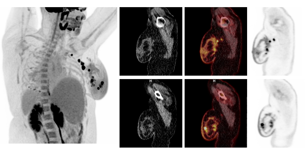
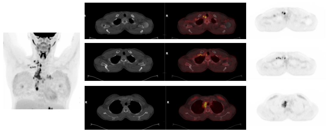
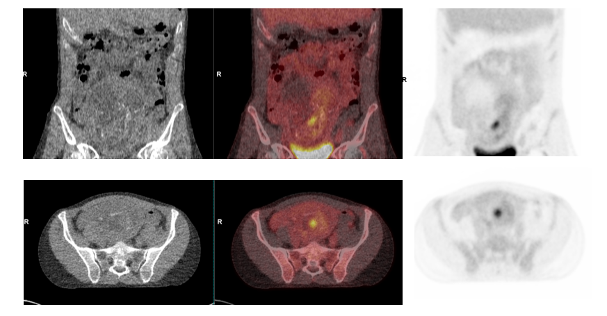
As shown in Figures 5 and 6, 18F-FDG PET/CT shows multiple moderate to intensely avid upper thoracic and mediastinal lymph nodal lesions involving right supraclavicular, right paratracheal, and right and left hilar, with the largest lesion in the right hilum measuring 39x24 mm with SUVmax = 9.7. The largest upper paratracheal node measured 1.4 cm in diameter with SUVmax = 8.3. The right epicardiac node shown in Figure 5 had SUVmax = 10.5, which is the highest among thoracic nodal lesions.
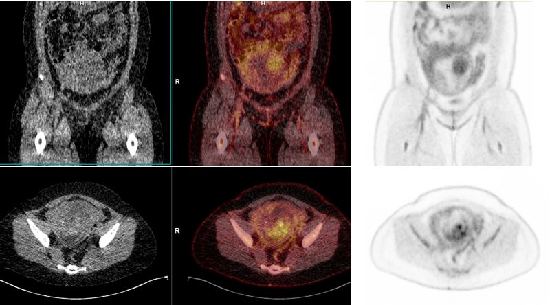
No 18F-FDG-avid nodes were visualized below the diaphragm. Bone marrow uptake was within normal limits. Liver and splenic uptake and size within normal limits. No hypermetabolic extra-lymphatic sites were visualized.
Bilateral breast prostheses visualized with mild diffuse bilateral breast tissue uptake reflecting pregnancy related proliferation.
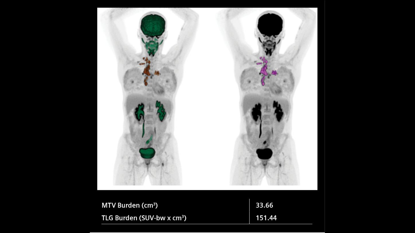
The study was consistent with extensive lymphomatous involvement in mediastinal and supraclavicular nodal groups but without involvement of any other distinct nodal groups including bilateral axillary or nodes on the other side of diaphragm. Total tumor burden and metabolic tumor volume estimations (Figure 7) were performed as a benchmark for post therapy evaluation of response.
High overall image quality with high lesion contrast in the lymph nodal lesions was obtained with 20-minute head-to-mid-thigh PET/CT study despite the very low injected dose due to the high sensitivity of the Biograph Vision Quadra with ultrafast time of flight capability and 106 cm FOV.
* Based on bench testing (e.g., improved sensitivity and ToF per NEMA NU-2:2018). Please refer to the approved PET drug prescribing information for dosing and administration instructions.
Case 2
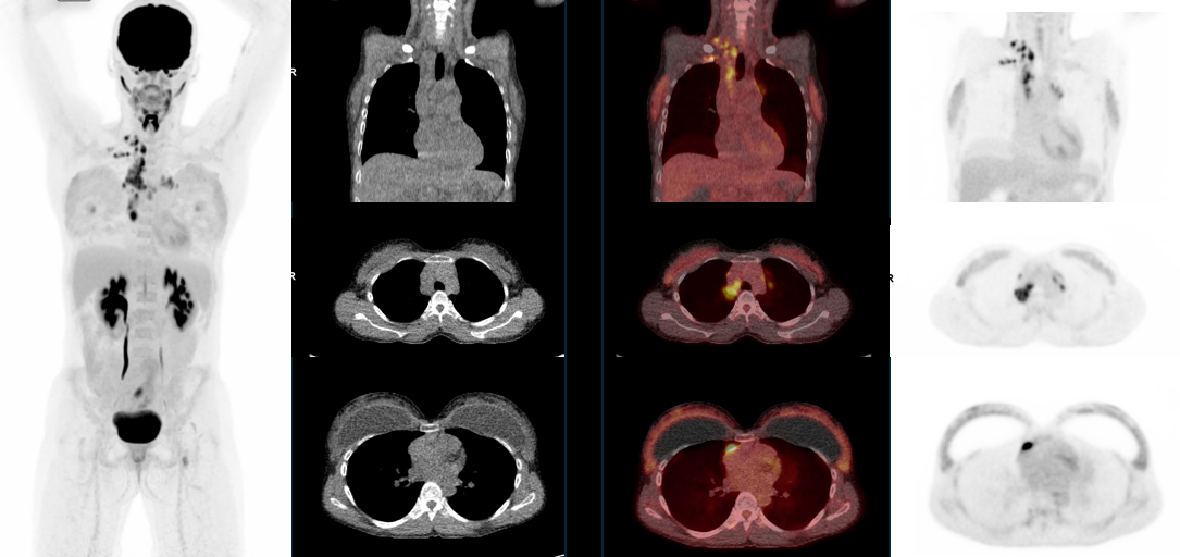
Figure 5: PET MIP and coronal and axial CT, PET, and fused PET/CT images at the hilar and lower thoracic level show 18F-FDG-avid enlarged mediastinal lymph nodal lesions involving right and left hilar and right paratracheal and anterior paracardiac lymph nodes. Coronal images also reveal multiple upper mediastinal lymph nodes.
Discussion
These two cases illustrate the capability of the Biograph Vision Quadra system to deliver high quality PET/CT imaging with extremely low injected dose without unduly increased acquisition time. The 15- and 20-minute scan times for head-to-mid-thigh acquisition in a single bed position with injected dose of approximately 40-60 MBq (1-1.5 mCi) achieved high image quality within acquisition times, which were acceptable to the departmental workflow and reduced undue patient motion. The high lesion contrast evident from the PET images and the SUVmax values reflects the high count statistics and high resolution of the 3.2 mm x 3.2 mm LSO detectors, as well as the ultrafast time of flight performance of the system. Generating high quality images at extremely low dose opens the possibility of imaging pregnant patients without serious risk of excessive fetal radiation dose and performing routine low dose evaluation of at-risk populations.
In a recent study from University Medical Center Groningen,4 10 pregnant patients were evaluated on the Biograph Vision Quadra with approximately 20 MBq 18F-FDG injection with 15-minute head-to-mid-thigh single bed position acquisition. All patients reported excellent image quality. A simulation study was performed to determine if the injected dose could be lowered while still achieving acceptable image quality without increasing scan time. The study showed that for a 15-minute scan time on the Biograph Vision Quadra, the injected dose could be reduced by around 40% without losing any clinical diagnostic confidence. The fetal dose calculated from an approximately 20 MBq 18F-FDG dose was 0.18 mGy, while the low dose CT with kVp Sn 140 and reference mAs of 10 delivered a fetal dose of 0.3 mGy, which together was deemed very low.
Using very low dose CT for attenuation correction is also key for fetal dose reduction. The Sn filter available in the Biograph Vision Quadra can absorb lower energy photons from the x-ray beam and can reduce radiation dose by around 25% in adults with BMI around 25 and up to 45% in adults with BMI>40. This was recently demonstrated in a series of obese patients from MD Anderson Medical Center.5
Conclusion
The combination of high sensitivity PET acquisition and ULD CT with Sn filter makes the Biograph Vision Quadra ideal for achieving high image quality with very low injected dose. This opens possibilities for imaging a wide range of patient populations including pregnant patients and pediatric patients with high radiosensitivity, as well as for patients with a high risk of cancer.
Examination protocol: Case 1
Scanner: Biograph Vision Quadra
Examination protocol: Case 2
Scanner: Biograph Vision Quadra
Prescribing information
Please see Indications and Important Safety Information.