
Physicians outside hospital imaging departments may not always recognize the potential benefits nuclear medicine and molecular imaging offer. Given the ever-changing healthcare landscape—one that is now even more fluid and volatile since the arrival of SARS-CoV-2—education and collaboration between nuclear medicine professionals and their colleagues are key to expanding and optimizing nuclear medicine’s utilization.
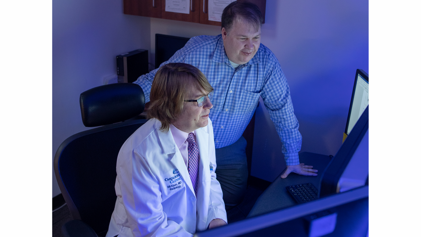
At Carilion Roanoke Memorial Hospital (CRMH), a community hospital serving Roanoke and the surrounding areas in Virginia, USA, Jackson W. Kiser, MD, section chief for molecular imaging, and James R. Crowley, MHA, CNMT, imaging services manager for nuclear medicine, PET/CT, and CT, have been working together since 2015 to ensure their department offers the most precise and effective nuclear medicine practices. They focus on building relationships with specialists and surgeons in other departments by educating them about the benefits of new approaches, not only in diagnostics, but in disease treatment and management. “We typically talk about evidence-based practices first, improved outcomes second, and cost much later,” Crowley explains.
Kiser and Crowley describe their work promoting underutilized areas of nuclear medicine as akin to being door-to-door salesmen. “Hospital physicians aren’t familiar with imaging; it’s our specialty and we need to teach them how to use it to the benefit of their patients,” states Kiser. Educational and collaborative efforts are the hallmark of how Kiser and Crowley navigate the changing healthcare landscape and strategically guide their practice to offer imaging solutions that aid in the best care delivery to patients at CRMH.
Switching cardiac imaging from SPECT to PET
To ensure CRMH offers the best cardiac-imaging practice, Kiser and Crowley focus their efforts on moving certain cardiac examinations from SPECT to PET.
Both had previous experience with cardiac PET before they joined CRMH, “so with some coaxing, we were able to convince our colleagues it was a better way to go,” Crowley recalls. The decision was supported by a powerful statement issued in 2016 by the American Society of Nuclear Cardiology (ASNC) and the Society of Nuclear Medicine and Molecular Imaging (SNMMI) that detailed the recommendations for myocardial perfusion PET based on its superior performance over other noninvasive diagnostic methodologies.1
Crowley stresses that from the administrator’s point of view, the major advantages of cardiac PET, when compared to nuclear cardiac examinations, are a decreased length of stay and elimination of two-day procedures. “A typical nuclear cardiac exam on an inpatient would take between three and five hours to get quality images, whereas cardiac PET can take 30 minutes. Additionally, inpatient nuclear cardiac exams are challenging due to confounding issues such as general inactivity, lack of bowel motility, increased bowel uptake, and general malaise, which leads to less treadmill and more chemical stress testing and repeat exams,” he notes. Accompanying an increase in the utilization of cardiac PET comes a shift in cardiac PET tracers, in which Kiser anticipates, “we will retain rubidium [82Rb] but we expect to eventually move to ammonia [13N].” CRMH is planning a large prospective, randomized study to compare the two radioisotopes in around 1,000 patients.
“By utilizing cardiac PET we reduce the number of unnecessary invasive procedures, the need for catheterization, patient length of stay, and we are able to scan more patients.”

Navigating change with education and collaboration
As with many changes, there was some initial resistance regarding the switch to cardiac PET among hospital cardiologists, “but once they learned how to read the scans, they embraced it,” Kiser and Crowley recollect. “We were lucky to be able to make the switch collegially,” Kiser stresses. “My counterpart in cardiology, Dr. Matthew Schumaecker, had experience with cardiac PET and knew its power.” Crowley pays tribute to the collaboration between Kiser and Schumaecker for their ability to build the bridge between their two departments, and notes, “it’s a very important aspect of why we have been successful in this effort.”
Concerns about costs associated with PET sometimes arise, but Kiser stresses the necessity of examining the cost to the overall facility, not just one department. “By utilizing cardiac PET we reduce the number of unnecessary invasive procedures, the need for catheterization, patient length of stay, and we are able to scan more patients. We believe the overall cost to the facility is comparable to that of cardiac SPECT.”

Expanding SPECT imaging in non-cardiac areas
Kiser and Crowley recognized that moving cardiac imaging to PET would create a deficiency in SPECT procedures, since cardiac imaging comprised 40% of the total volume of nuclear medicine procedures, which translated to a 40% reduction in total billable procedures in the nuclear medicine department. Today, those 40% of SPECT cardiac procedures have been replaced with other types of SPECT examinations. “We started doing SPECT/CT whenever we needed to better delineate anatomy,” Crowley recalls. “We began with lymphoscintigraphy for our surgeons and then moved to diagnosis of gastrointestinal bleeding for our interventional radiologists.” They also identified new opportunities to utilize SPECT/CT in the evaluation of bone lesions caused by primary prostate and breast cancers. “We believed that blastic components would benefit from a more sensitive and accurate exam,” Kiser explains. “Now all these patients have a whole-body SPECT/CT.”
CRMH recognized the potential for SPECT/CT in orthopedics, particularly spine work. Ethan Colliver, DO, a specialist in physical medicine and rehabilitation, based nearby in Blacksburg, Virginia, was an early adopter.2 Since the hospital’s own spine surgeons never ordered nuclear exams, “we talked to them about SPECT/CT and began the conversation by detailing how SPECT/CT may be better clinically than MRI in some indications relating to physiology. We also explained SPECT/CT’s dose is comparable to, or less than, a CT scan,” Crowley relates.
Orthopedic studies account for 30 to 40 percent of SPECT examinations performed at the hospital, with the remaining procedures primarily attributed to oncology. Expanding beyond these two indications, CRMH has also moved SPECT utilization into neurology by conducting ictal SPECT examinations for epileptic patients. In the pulmonary field, V/Q lung SPECT/CT will be used for the visualization of suspected pulmonary embolism. “We will roll it out for emergency and in-house patients to determine how it impacts critical care and pulmonology practice, then start it for the cardiothoracic surgeons for preoperative lung resection planning,” Kiser anticipates.
Building bridges for future expansion
It’s apparent Kiser and Crowley are invested in preparing their department for the future.
Envisioning their practice moving forward, Kiser declares, “theranostics is the biggest thing in nuclear medicine right now and we want to be at the forefront. With theranostics you can eliminate unnecessary surgeries and use a targeted molecular therapy that increases overall survival by years. This approach to personalized medicine is the way of the future, and we need to embrace it and figure out how to do it better,” he says. To start, the hospital’s oncology and nuclear medicine departments collaborated and received approval to convert one of the hospital buildings into a theranostics center, which is scheduled to begin inpatient and outpatient services in 2020.
Kiser urges, “You can’t be a radiologist who just sits in the office waiting for images to arrive at your desk where you read them and push them out. You have to be interactive, proactive, and be an educator and advocate.”

Our response to the arrival of SARS-CoV-2
At the time of this interview, we were not yet fully aware of the impact SARS-CoV-2 would have on the healthcare community, and the far-reaching impacts of the virus may not be fully transparent for quite some time. The pandemic is unprecedented and how each healthcare facility, and individual department, must adapt is unprecedented as well.
At Carilion Roanoke Memorial Hospital, our nuclear medicine department is no exception. Like many other fields, we found ourselves in a situation where, in order to adapt adequately, we had to prioritize. We began by reviewing all examination requests to determine what should be deemed more urgent than others. We decided studies that were not urgent or emergent would be postponed for 30 days and reassessed at that time, after the immediate contingency plans to run the hospital were put in place. Then, once there was an additional plan to safely manage and navigate patients through potential COVID-19 areas, non-emergent scans could resume. Examples of these types of patients were gastric emptying scans, renograms, and gallbladder scans that were not for acute cholecystitis.
All scans for patients being staged, restaged, or undergoing treatment monitoring for cancer were kept on schedule. And, of course, any emergent or urgent scans remained a first priority, such as ventilation/perfusion (V/Q) for pulmonary embolism, myocardial perfusion, and gastrointestinal bleeding.
Our approach to the crisis was, and continues to be, one based in common sense; once the process established at our facility was fully functional, we learned to operate within a new normal. But our need to adapt is far from over, we still have to work to define the parameters of our practice’s new normal: what is an acceptable volume, an acceptable amount of patients, and how we will continue to operate and define “new normal” as the situation progresses? It’s an evolving target.
About the author
Linda Brookes is a freelance medical writer and editor who divides her time between London and New York, working for a variety of clients in the healthcare and pharmaceutical fields.
