Holger Nef, MD, is one of the world’s leading interventional cardiologists. Besides relying on established and innovative intravascular imaging procedures and noninvasive ischemia testing solutions, he also advocates combining all available relevant information to create a comprehensive clinical picture of the patient. The aim is to enhance the quality of treatment in the long term. In this interview, the associate director of the University Hospital of Giessen and Marburg’s Cardiovascular Center discusses trends in interventional cardiology, the strengths and weaknesses of intravascular imaging, and his ideas for training new professionals.
Photos: Sebastian Bullinger
Download your print version here.

Dr. Nef, in your view, how has cardiology changed in recent years?
Nef: The field of medicine in general has been heavily influenced by growing cost pressure on the one hand, and rising quality standards on the other. Striking the right balance between these two factors is the name of the game right now. In regards to cardiology, I think the most striking change is the succession of technological developments we have seen in recent years: For instance, whereas in 2008 TAVI was still in its infancy, it has now become a standard procedure, performed some 17,000 times a year in Germany. Or think of percutaneous AV treatments, where major developments have occurred at a very fast pace. We are now able to perform a mitral valve annuloplasty by applying a band in a way that emulates certain aspects of cardiac surgical procedures, enabling us to provide treatment in almost the same way as cardiac surgeons, but in a noninvasive manner.
How have diagnosis, treatment, and follow-up of coronary heart disease changed in this period?
Nef: In my opinion, the key changes concerning diagnosis of coronary heart disease relate to the functional measurement of stenosis. The ability to measure functional flow reserve is a major step forward in the detection of coronary stenosis requiring treatment. As regards the treatment of coronary heart diseases, we have seen substantial improvements above all in catheter and stent systems.
Developments in stents are not limited to material changes; also notable is the use of narrower stent struts. This has led to significant improvements in our patients’ clinical outcomes. Nevertheless, this has not completely eliminated the risks arising from implantation, so we were naturally very excited about the development of bioresorbable scaffolds.
However, as a profession, we were not aware that these new scaffolds called for special techniques during implantation in order to ensure a positive long-term outcome. Accordingly, the latest available results are somewhat discouraging. That said, an improved version of the scaffolds that could potentially replace metal in future is currently being tested.
“My own experience shows that intracoronary imaging allows us to perform stent implants much more safely and efficiently.”
How do you assess your hospital’s position in the field of diagnostic imaging?
Nef: For a university cardiac center like ours in Giessen, it is crucial to be able to offer state-of the- art diagnostic imaging technology. We were among the first centers in Germany to acquire the advanced computed tomography system SOMATOM Force. This meant we could offer our patients a particularly mild form of coronary CT diagnosis at a very early stage. Besides providing a reliable way to rule out coronary heart disease, this technology can also be used to verify treatment results in certain cases. Meanwhile, the latest developments and systems in the field of cardiac MRI can answer the questions regarding surrounding myocardial ischemia/disease with a high degree of precision.

What can you gain from superimposing ultrasound and fluoroscopy images?
Nef: We have only been using the new syngo TrueFusion imaging technology in clinical practice for a short time. Nevertheless, we can already say that the combined information from echocardiography and fluoroscopy gives us precisely the anatomical details that we need to position our implants even more accurately and safely. For the new mitral valve annuloplasty procedure in particular, we gain valuable additional information that can considerably reduce examination times – and therefore radiation exposure for both patients and examining physicians.
What specific challenges do you see in noninvasive CT imaging?
Nef: The great strength of CT imaging lies in its specificity. Yet, assessing the extent of a stenosis on the basis of plaque morphology remains difficult. In such cases, divergent findings are still very common, but that does not diminish the huge predictive value of a CT scan. And our goal is quick, early rule-out of coronary heart disease in patients with medium pretest probability. A significant step forward will be noninvasive flow measurement, which will help distinguish a relevant coronary stenosis from an insignificant one.
How important is the issue of dose reduction in your work, and how do you implement the ALARA principle?
Nef: This is an extremely important issue for us. These days, we always begin our X-ray-based examinations with low-dose scans, and are often amazed at how high the image quality is, even with the lowest possible dose. Nevertheless, some examinations naturally require a higher level of radiation. The main thing is that we learn to work with as little radiation as possible, and I think that Siemens Healthineers provides us with effective support in this regard.
“I think that we should no longer implant stents at all without prior ischemia testing. Not just because our guidelines say so, but because it leads to demonstrably higher-quality results.”
You have some experience with stent enhancement solutions such as CLEARstent. What benefits do you think these technologies have to offer?
Nef: The focus of these technologies is on enabling better stent positioning. Stent enhancement solutions help prevent inaccurate stent positioning. Furthermore, they allow us to assess the overall expansion of the stent. I like to compare CLEARstent to assisted parking technology: It is incredibly helpful, but of course it can’t completely replace looking over your shoulder. In short, I am an avid user of stent enhancement technologies, and I think they can help us position stents with greater overall safety and accuracy, which is obviously a good thing for our patients.

What are the benefits of intracoronary imaging in your view?
Nef: I think that procedures such as optical coherence tomography (OCT) or intravascular ultrasound (IVUS) offer enormous benefits, at least as far as evaluation of plaque morphology and strategy planning are concerned. Here, too, there is a shortage of good randomized studies on actual patient outcomes. But my own experience shows that intracoronary imaging allows us to perform stent implants much more safely and efficiently, because we know immediately after placing the stent whether it is properly positioned and whether any vessel damage has occurred. In clinical practice, we use OCT and IVUS in a complementary fashion, as both methods have their strengths and weaknesses. OCT offers significantly higher resolution than IVUS, but requires a contrast agent, which limits usability in patients with renal insufficiency. Furthermore, it only offers a penetration depth of around 2 mm, making it less suitable for larger vessels. In other words, we use OCT wherever possible, and in all other cases we are glad to be able to fall back on IVUS.

Do you make use of the possibilities arising from co-registration of angiography and OCT?
Nef: Absolutely. This option has made OCT even safer, as angiography gives us the precise location of our OCT catheter at all times. Furthermore, by combining the two images we can evaluate the lesion much more accurately than without co-registration.
What do you think of the potential of noninvasive FFR measurement?
Nef: I think that we should no longer implant stents at all without prior ischemia testing. Not just because our guidelines say so, but because it leads to demonstrably higher-quality results. This is supported above all by the results of the FAME 2 study. I think it would be desirable in future to be able to perform the steps to determine FFR in parallel and in a largely automated manner – this would give us a kind of live analysis, supporting faster decision-making.
What developments do you anticipate in the field of diagnostic imaging?
Nef: In future, there will be less of a focus on imaging and much more on information itself. Big Data is a concept I am not particularly fond of, but it neatly captures where the field is headed. In future, we will collect and automatically analyze images, medical history, lab data, and information on cardiac function and previous conditions. New systems will suggest treatment methods or offer strategy recommendations for us to adapt to the patient setup. I think this will also present a major opportunity for interdisciplinary cooperation: Putting the patient at the center and connecting the various dots to form a comprehensive overall picture will substantially facilitate decision-making in everyday practice.
What are your expectations of an industry partner like Siemens Healthineers for future technological developments?
Nef: I don’t think that developers, engineers, and medical practitioners can cooperate closely enough for our practical needs to be implemented in products and solutions. Take the continued fusion of intravascular imaging with fluoroscopy, for example. Here we need solutions for automatic transmission of stent dimensions onto the angiography image so that the stent marker can be automatically recognized and the system can tell me when the stent is in the right position. Another issue is training new professionals. I think there is a huge need for simulation tools on which interventions can be practiced. A trainee pilot doesn’t simply sit behind the controls of an A380, take off in San Francisco then land in Hong Kong. I believe that the field of medicine also needs good simulation-based training programs to give young professionals the best possible preparation for the huge responsibilities they are to be entrusted with.

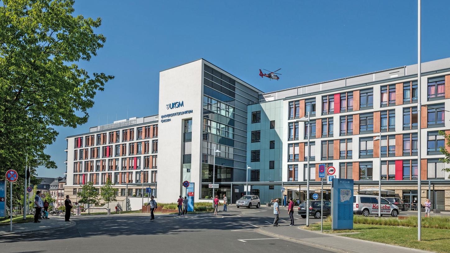
The University Hospital of Giessen and Marburg, part of the Rhön Klinikum AG hospital group, was formed by the merger between the university hospitals of Justus Liebig University Giessen and Philipps University Marburg. The Cardiology and Angiology Clinic at the Giessen site alone performs more than 4,000 catheter-based interventions a year.


