
ISMRM & SMRT Virtual Conference & Exhibition 2021Exploring new frontiers in MRI, together.
Visit Siemens Healthineers at our virtual booth
Exploring new frontiers in Precision Medicine
Join us at this year´s virtual ISMRM and discover how our solutions constantly exceed the possible. The innovative spirit of this unique, vibrant MRI community really comes to life at ISMRM and Siemens Healthineers is proud to be part of this network. The passion for jointly pioneering MRI around the globe is truly unmatched.
Lunch Symposium
To experience the latest MRI developments and gain insights into future innovations, please join our Siemens Healthineers MRI Symposium at the ISMRM 2021.
Time:
Tuesday, May 18, 2021, 10:00 -
11:00 UTC
Location:
The Siemens Healthineers MRI Symposium will be available on the platform of ISMRM & SMRT Virtual Conference & Exhibition.
Speaker
- Welcome
Christiane Bernhardt
VP Marketing & Sales Magnetic Resonance, Siemens Healthineers - Opportunities at 0.55T
Prof. Krishna Nayak
University of Southern California, USA - Project to develop a next generation 7T MRI brain scanner for high resolution imaging
Prof. David Feinberg
University of California - Berkeley, USA - Clinical applications of UHF MRI - a first year's experience
Prof. Dr. Roland Wiest
Inselspital Bern, Switzerland - The connected imaging instrument
Dr. Michael Hansen
Microsoft Corporation, Washington, USA
Highlights

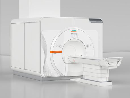
MAGNETOM Free.Max1 – Breaking barriers
MAGNETOM Free.Max breaks barriers to expand the reach of MRI. Where patients have felt discomfort, the world's first 80 cm bore sets a new paradigm in patient comfort. Where infrastructure was an obstacle to MRI, MAGNETOM Free.Max slots into an existing helium-free infrastructure. Where access to MRI was not viable, MAGNETOM Free.Max makes access affordable. And where conventions have limited our thinking, MAGNETOM Free.Max breaks out of conventions to explore new clinical opportunities in MRI.

MR Fingerprinting2 – A giant leap for precision medicine
What if we could unlock the power of quantitative data in MR to improve diagnostic accuracy? Magnetic Resonance Fingerprinting (MRF)2 uses quantified information to generate a more precise under¬standing of a patient’s condition. MRF has enormous potential to improve tissue differentiation and enable less invasive diagnostics. Reliable MRF data could increase objective comparisons in follow-up studies. Ultimately, aided by AI, quantitative measurements could lead to more personalized treatment. MRF is at the frontier of a new dimension in quantitative imaging.
MAGNETOM Terra – Translate 7T research power into clinical care
Explore new territories in MRI by enabling powerful 7T research and enhancing clinical care and better patient outcomes. MAGNETOM Terra is the first 7T scanner released for clinical use in Europe and US. The unique Dual Mode lets you switch between clinical and research operations, with separate database to distinguish between clinical and research scans. The advanced ultra-high-field (UHF) technology has the power, to put you firmly at the forefront of MRI – giving you a competitive edge and increasing your reputation as a true opinion leader.

Deep Resolve – Mobilizing the power of networks

AI-Rad Companion
The AI-Rad Companion, our family of AI-powered, cloud-based augmented workflow solutions, helps you to reduce the burden of basic repetitive tasks and may increase your diagnostic precision when interpreting medical images.
Its solutions provide automatic post-processing of imaging datasets through our AI-powered algorithms. For example, the AI-Rad Companion assists in the interpretation of MR images of the brain. AI-Rad Companion Brain MR for morphometry analysis automatically segments different brain areas and quantifies each of their volumes. These results are compared to a normative database and can easily be assessed by the radiologist through a color-coded deviation map and a quantitative overview. The AI-Rad Companion portfolio includes further offerings that use MRI images for AI-based image quantification. AI-Rad Companion Prostate MR for biopsy support automatically segments the prostate and allows the manual annotations of lesions, which can support in targeted biopsies under MRI and Ultrasound fusion imaging. Targeted, MRI-supported biopsies like this can make it easier for the urologist to detect significant prostate carcinomas and improve the quality of patient care.
The product is pending 510(k) clearance and is not yet commercially available in the United States. Its future availability cannot be guaranteed.